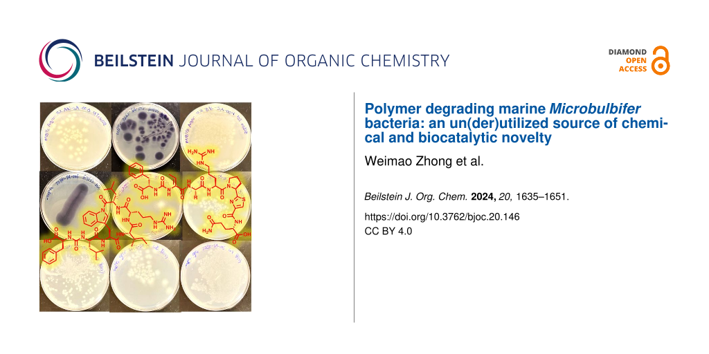
Abstract
Microbulbifer is a genus of halophilic bacteria that are commonly detected in the commensal marine microbiomes. These bacteria have been recognized for their ability to degrade polysaccharides and other polymeric materials. Increasingly, Microbulbifer genomes indicate these bacteria to be an untapped reservoir for novel natural product discovery and biosynthetic novelty. In this review, we summarize the distribution of Microbulbifer bacteria, activities of the various polymer degrading enzymes that these bacteria produce, and an up-to-date summary of the natural products that have been isolated from Microbulbifer strains. We argue that these bacteria have been hiding in plain sight, and contemporary efforts into their genome and metabolome mining are going to lead to a proliferation of Microbulbifer-derived natural products in the future. We also describe, where possible, the ecological interactions of these bacteria in marine microbiomes.

Graphical Abstract
Introduction
Microbial natural products represent an important reservoir for drug discovery and along with their (semi)synthetic derivatives, constitute a major component of the contemporary pharmaceutical arsenal. Their thusly validated utility is juxtaposed against the barriers to natural product discovery; high rediscovery rates, reliance on largely serendipitous response in bioactivity assays, and the resources and expertise required for their structure determination being the primary impediments [1,2]. With this background, accessing biological sources that have not yet been extensively mined for natural product discovery is a promising route, one which, at the very least, promises to ameliorate the problem of continued rediscovery of known natural products. This review outlines the recent progress that has been realized using bacteria of the genus Microbulbifer. Together with the emerging field of natural product discovery from Microbulbifer bacteria, this genus of obligate marine bacteria is well validated to be a valuable source of biopolymer degrading enzymes. In this review, we outline the discovery of Microbulbifer enzymes that have substantial biocatalytic potential as it pertains to biopolymer degradation, followed by a description of natural product discovery from this genus.
Microbulbifer is a genus of rod-shaped, aerobic Gram-negative Alteromonadaceae family γ-proteobacteria. They are halophilic obligate marine bacteria that are frequently isolated from diverse marine holobionts such as sponges [3,4], sediments [5], algae [6], and corals [7] using media that mimic the native salt water habitat. Strains of this genus are detected together with Pseudovibrio and Ruegeria strains that grow in identical culture conditions, and hence obtaining axenic Microbulbifer strains may require repeated rounds of serial enrichment. This genus was first described by Gonzales et al. in 1997 [5]. More than 130 strains have been reported from regions of wide geographical dispersion (Table 1, Figure 1). Microbulbifer bacteria are well known for their capacity to degrade not only a wide variety of polysaccharides including cellulose, agar, chitin, alginate, and xylan [8-11], but also plastics [4,12]. Progressing from their isolation and cultivation, natural product discovery from Microbulbifer bacteria is starting to gather steam [6,13,14]. The major classes of natural products isolated from Microbulbifer to date include alkaloids [15], fatty acid and polyketides [16,17], and non-ribosomally synthesized peptides [3,4,7]. These findings validate the potential for secondary metabolite discovery from Microbulbifer genus. This review will cover the distributions and origins of Microbulbifer bacteria strains, degradation enzymes, and secondary metabolite discoveries. A focus is placed on the novel chemical structures reported with reference to their biological activities and the biosynthetic studies they have inspired.
Table 1: Summary of marine sources, geographical locations, and key characteristics of Microbulbifer strains reported in literature.
| Origin | Location | Key characteristics | References | ||
| 1 | Microbulbifer hydrolyticus gen. nov. | marine pulp mill | USA |
produces hydrolytic enzymes,
polyethylene degradation |
[5,18,19] |
| 2 | Microbulbifer degradans. 2-40 | salt marsh | USA |
degrades polysaccharides,
polyserine linkers, chitinase |
[20-22] |
| 3 | Microbulbifer salipaludis sp. nov. | salt marsh | Korea | –a | [8] |
| 4 | Microbulbifer arenaceous sp. nov. | red sandstone | Scotland | hydrolyzes chitin, esculin, gelatin, and starch | [23] |
| 5 | Microbulbifer elongates comb. nov. | –a | Germany | –a | [24] |
| 6 | Microbulbifer sp. JAMB-A7 | sediment | Japan | β-agarase | [25] |
| 7 | Microbulbifer sp. JAMB-A94 | sediment | Japan | β-agarase, ι-carrageenase | [10,26-29] |
| 8 | Microbulbifer sp. JAMB-A3 | sediment | Japan | β-agarase | [10,30] |
| 9 | Microbulbifer maritimus sp. nov. | sediment | Korea | –a | [31] |
| 10 | Microbulbifer sp. A4B-17 | ascidian | Palau |
produces 4-HBA and
parabens |
[6] |
| 11 | Microbulbifer sp. A4A-72 | sponge | Palau | produces 4-HBA | [6] |
| 12 | Microbulbifer sp. V296 | sponge | Yap | produces 4-HBA | [6] |
| 13 | Microbulbifer sp. A4B-20 | algae | Palau | produces 4-HBA | [6] |
| 14 | Microbulbifer sp. A2C-113 | sponge | Palau | produces 4-HBA | [6] |
| 15 | Microbulbifer sp. A4A-79 | ascidian | Palau | produces 4-HBA | [6] |
| 16 | Microbulbifer sp. AM222 | sediment | Palau | produces 4-HBA | [6] |
| 17 | Microbulbifer sp. AM292 | sediment | Palau | produces 4-HBA | [6] |
| 18 | Microbulbifer sp. A4A-68 | sponge | Palau | produces 4-HBA | [6] |
| 19 | Microbulbifer sp. A2D-17 | sponge | Palau | produces 4-HBA | [6] |
| 20 | Microbulbifer sp. I1876 | algae | Japan | produces 4-HBA | [6] |
| 21 | Microbulbifer sp. C91 | algae | Yap | produces 4-HBA | [6] |
| 22 | Microbulbifer sp. HG111 | sponge | Japan | produces 4-HBA | [6] |
| 23 | Microbulbifer sp. F-104 | algae | Palau | produces 4-HBA | [6] |
| 24 | Microbulbifer sp. V32 | sponge | Yap | produces 4-HBA | [6] |
| 25 | Microbulbifer sp. HG125 | sponge | Japan | produces 4-HBA | [6] |
| 26 | Microbulbifer sp. A3G-2 | ascidian | Palau | produces 4-HBA | [6] |
| 27 | Microbulbifer sp. A3P2-23-3-2 | ascidian | Palau | produces 4-HBA | [6] |
| 28 | Microbulbifer sp. AM220 | sediment | Japan | produces 4-HBA | [6] |
| 29 | Microbulbifer sp. YM3-0188 | sponge | Palau | produces 4-HBA | [6] |
| 30 | Microbulbifer sp. A2D-15 | sediment | Palau | produces 4-HBA | [6] |
| 31 | Microbulbifer sp. HG869 | ascidian | Palau | produces 4-HBA | [6] |
| 32 | Microbulbifer sp. HG279 | sponge | Japan | produces 4-HBA | [6] |
| 33 | Microbulbifer sp. ssthio04PA2-36a | sponge | Palau | produces 4-HBA | [6] |
| 34 | Microbulbifer celer sp. nov. | saltern | Korea | –a | [32,33] |
| 35 | Microbulbifer halophilus sp. nov. | saline soil | China | –a | [34] |
| 36 | Microbulbifer sp. CMC-5 | seaweed | India |
degrades polysaccharides,
β-agarase |
[35-37] |
| 37 | Microbulbifer variabilis sp. nov. | algae | Japan | –a | [38] |
| 38 | Microbulbifer epialgicus sp. nov. | algae | Japan | –a | [38] |
| 39 | Microbulbifer sp. L4-n2 | sponge | France | produces parabens | [13] |
| 40 | Microbulbifer donghaiensis sp. nov. | sediment | China | –a | [39] |
| 41 | Microbulbifer chitinilyticus sp. nov. | mangrove | Japan | chitin-degrading | [9] |
| 42 | Microbulbifer okinawensis sp. nov. | mangrove | Japan | chitin-degrading | [9] |
| 43 | Microbulbifer sp. SD-1 | seawater | Korea | degrades agar | [40] |
| 44 | Microbulbifer sp. JAM-3301 | sediment | Japan | inulinase producer | [41] |
| 45 | Microbulbifer maritimus | seaweed | India | agarase producer | [42] |
| 46 | Microbulbifer sp. 6532A | seaweed | Japan | alginate lyase producer | [43,44] |
| 47 | Microbulbifer marinus sp. nov. | sediment | China | –a | [45] |
| 48 | Microbulbifer yueqingensis sp. nov. | sediment | China | –a | [45] |
| 49 | Microbulbifer gwangyangensis sp. nov. | tidal flat | Korea | –a | [46] |
| 50 | Microbulbifer pacificus sp. nov. | sponge | Korea | –a | [46] |
| 51 | Microbulbifer mangrove sp. nov. | mangrove | India | polysaccharide-degrading | [11] |
| 52 | Microbulbifer elongatus HZ11 | seawater | China | seaweed-degrading | [47] |
| 53 | Microbulbifer thermotolerans DAU221 | sediment | Korea |
produces carbohydrate esterase, α-amylase, esterase, chitinase, GH3
-glucosidase polysaccharide lyase |
[48-53] |
| 54 | Microbulbifer elongatus sp. A13 | seaweeds | India | agarase producer | [54] |
| 55 | Microbulbifer rhizosphaerae sp. nov. | halophytic plant | Spain | –a | [55,56] |
| 56 | Microbulbifer sp. ALW1 | algae | China | produces alginate lyase, laminarinase, β-glucosidase, chondroitinase | [12,57-62] |
| 57 | Microbulbifer flavimaris sp. WRN-8 | sediment | China | –a | [63] |
| 58 | Microbulbifer mangrovistrain DD-13 | mangrove | India | degrades polysaccharides | [64] |
| 59 | Microbulbifer echini sp. AM134 | sea urchin | Korea | –a | [65] |
| 60 | Microbulbifer aggregans sp. CCB-MM1 | sediment | Malaysia | produces sulfite reductase | [66-68] |
| 61 | Microbulbifer aestuariivivens sp. GHTF-23 | tidal flat | Korea | –a | [69] |
| 62 | Microbulbifer sp. Q7 | sea cucumber | China | produces β-agarase, alginate lyases | [70-72] |
| 63 | Microbulbifer sp. 127CP7-12 | sponge | Korea | produces violacein | [73] |
| 64 | Microbulbifer hydrolyticus IRE-31-192 | chemical mutation | China | polyethylene degradation | [74] |
| 65 | Microbulbifer sp. GL-2 | blackfish | Japan | cellulase | [75] |
| 66 | Microbulbifer agarilyticus. GP101 | invertebrate turbo cornutus | Korea | –a | [76] |
| 67 | Microbulbifer sp. BN3 | sediment | China | β-agarase, chitinase | [77-79] |
| 68 | Microbulbifer sp. WMMC-695 | tunicate | USA | produces antibacterial acetamido-4-hydroxybenzoate esters | [14] |
| 69 | Microbulbifer sp. BH-1 | seawater soil | China | β-agarase | [80] |
| 70 | Microbulbifer sp. C4-6 | coral | Japan | produces fatty acid with weak growth inhibition against Saccharomyces cerevisiae | [16] |
| 71 | Microbulbifer sp. AG1 | mangrove soil | China | β-agarase producer | [81] |
| 72 | Microbulbifer sp. KIT−19 | seawater | Japan | alginate lyase producer | [82] |
| 73 | Microbulbifer harenosus sp. nov. | coastal sand | China | alginate-degrading | [83] |
| 74 | Microbulbifer sp. DC3-6 | coral | Japan | produces antimicrobial and cytotoxic alkanoyl imidazoles | [15] |
| 75 | Microbulbifer sp. SH-1 | coastal soil | China | alginate lyase producer | [84] |
| 76 | Microbulbifer elongates. PORT2 | seawater | Indonesia | agarase producer | [85,86] |
| 77 | Microbulbifer hainanensis sp. nov. | sediment | China | –a | [87] |
| 78 | Microbulbifer sp. YNDZ01 | macroalgae | Indonesia | lipase, ι-carrageenase | [88,89] |
| 79 | Microbulbifer sp. CL37 | sediment | Malaysia | xylanase producer | [90] |
| 80 | Microbulbifer sp. SOL66 | soil | Korea | degrades poly(3-hydroxybutyrate) | [91,92] |
| 81 | Microbulbifer sp. SOL03 | soil | Korea | degrades poly(3-hydroxybutyrate) | [91,93] |
| 82 | Microbulbifer sp. SOL51 | soil | Korea | degrades poly(3-hydroxybutyrate) | [91] |
| 83 | Microbulbifer sp. SOL55 | soil | Korea | degrades poly(3-hydroxybutyrate) | [91] |
| 84 | Microbulbifer sp. SOL84 | soil | Korea | degrades poly(3-hydroxybutyrate) | [91] |
| 85 | Microbulbifer sp. GL-1 | blackfish | Japan | cellulase-producing | [94] |
| 86 | Microbulbifer sp. GL-2 | blackfish | Japan | cellulase-producing | [94] |
| 87 | Microbulbifer sp. GL-3 | blackfish | Japan | cellulase-producing | [94] |
| 88 | Microbulbifer sp. YPW1 | sediments | China | –a | [95] |
| 89 | Microbulbifer sp. SH-1 | soil | China | alginate lyase producer | [96] |
| 90 | Microbulbifer okhotskensis sp. КMM 9862 | sediment | Russia | –a | [97] |
| 91 | Microbulbifer sp. BY17 | gracilaria | China | alginate lyase producer | [98] |
| 92 | Microbulbifer sp. YX04 | mangrove | China | algicidal | [99] |
| 93–130 | Microbulbifer sp. | sponge | USA | –a | [100] |
| 131 | Microbulbifer sp. C10-1 | coral | Japan | produces cyclic hexapeptides | [7] |
| 132 | Microbulbifer sp. RZ01 | coastal environment | China | algicidal | [17] |
| 133 | Microbulbifer sp. TB12003 | coastal environment | China | nonalgicidal | [17] |
| 134 | Microbulbifer sp. MLAF003 | sponge | USA | produces cyclic hexapeptides | [4] |
| 135 | Microbulbifer sp. VAAF005 | sponge | USA | produces cyclic hexapeptides | [4] |
| 136 | Microbulbifer sp. MKSA007 | sponge | USA | produces linear peptides | [3] |
aNo information available.
![[1860-5397-20-146-1]](/bjoc/content/figures/1860-5397-20-146-1.png?scale=2.0&max-width=1024&background=FFFFFF)
Figure 1: Oceanic distribution and marine holobiont sources of Microbulbifer strains described in the literature.
Figure 1: Oceanic distribution and marine holobiont sources of Microbulbifer strains described in the literat...
Review
Biopolymer degrading enzymes from Microbulbifer
Agarases
Agarose is a natural polymer consisting of a linear chain of alternating residues of 3-O-linked β-ᴅ-galactopyranose and 4-O-linked 3,6-anhydro-α-ʟ-galactose. Agarose is a major component of agar, a polysaccharide present in the cell walls of some red algae [101]. Agarases are glycoside hydrolases (GHs) that degrade agarose to smaller oligosaccharides [102]. Marine bacteria of diverse genera produce agarases [103-105]. Based on the reactions that they catalyze, bacterial agarases are grouped as α- and β-agarases wherein they hydrolyze α-1,3 linkages and β-1,4 linkages in agarose, respectively (Figure 2). However, based on sequence similarity, agarases are alternatively classified into different families of glycoside hydrolases (GHs): GH-96 and GH-117 for α-agarases, and GH-16, GH-50, GH-86, and GH-118 for β-agarases [106]. All agarases characterized from Microbulbifer bacteria belong to β-agarases.
![[1860-5397-20-146-2]](/bjoc/content/figures/1860-5397-20-146-2.svg?scale=2.0&max-width=1024&background=FFFFFF)
Figure 2: The chemical structure of agarose with the key β-1,4 linkage denoted.
Figure 2: The chemical structure of agarose with the key β-1,4 linkage denoted.
GH-16 agarases: The first report for the characterization of an agarase from the Microbulbifer genus was from the strain Microbulbifer sp. JAMB-A7 isolated from deep-sea sediment [25] wherein the agarase encoding gene agaA7 was sequenced and cloned. The recombinant enzyme RagaA7 was produced in a Bacillus subtilis host and characterized to be a neoagarotetraose-producing GH-16 family endo-type β-agarase with a pH and temperature optima being 7.0 and 50 °C, respectively. Another thermostable neoagarotetraose-producing GH-16 endo-type β-agarase rAgaA was identified and cloned from deep-sea-derived Microbulbifer sp. JAMB-A94 with pH and temperature optima being 7.0 and 55 °C, respectively [26]. The recombinant enzyme was likewise produced using a B. subtilis host. The crystal structure of the catalytic domain was determined to show a β-jelly roll fold with structural similarity to two other β-agarases, ZgAgaA and ZgAgaB, from Zobellia galactanivorans, which is a model marine bacterium for the bioconversion of algal biomass [29,107-109]. The crystal structure suggested that the thermostability of rAgaA likely derives from the numerous surface salt bridges, relatively short length of the surface loops, and the increased number of Pro and Arg residues [29].
A truncated β-agarase gene without the carbohydrate-binding modules (Aga16A-ΔCBM) from the marine Microbulbifer sp. BH-1 was cloned and expressed in Escherichia coli with pH and temperature being 8.0 and 55 °C, respectively. The biocatalytic utility of the enzyme was also evaluated; it was used for neoagarooligosaccharide production in high yield [80].
Five additional GH-16 agarases AgaA3 produced in B. subtilis, ID2563 produced in E. coli, N3-1 produced in Pichia pastoris, AG1 produced in E. coli, and AgaF16A produced in E. coli, were cloned and expressed from marine Microbulbifer bacteria Microbulbifer sp. JAMB-A3 [30], Microbulbifer sp. Q7 [70], Microbulbifer sp. BN3 [77], Microbulbifer sp. AG1 [81], and Microbulbifer elongatus PORT2 [86], respectively. These agarases similarly degraded agarose into neoagarotetraose. With a view towards their biotechnological utility, AgaA3 was stable to high concentrations of surfactants.
GH-50 agarases: The strain Microbulbifer elongatus PORT2 was reported to be isolated in Indonesia [85]. In the sequenced genome of this bacterium were detected three agarases to be encoded which belonged to the GH-50 agarase family. The enzymes were named AgaA50, AgaB50, and AgaC50. Biochemical characterization of these three enzymes revealed that AgaA50 and AgaC50 generated neoagarobiose products implying cleavage within the agarose polymer (endolytic hydrolysis). The activity of AgaB50 was different, in that this enzyme cleaved at the external periphery of the agarose polymer to generate neagarosetetraose and neoagarobiose.
GH-86 agarases: The Microbulbifer sp. JAMB-A94 was shown to produce a GH-16 agarase (rAgaA) [26]. A gene encoding (agaO) for a novel GH-86 family agarase was also reported from the same bacterium [27]. The recombinant enzyme (rAgaO) was expressed in B. subtilis cells to demonstrate that this enzyme was an endo-type β-agarase and degraded agarose to neoagarohexaose as the main product [27]. The enzyme rAgaO represents the first and only report on the characterization of a purified agarase belonging to the family GH-86 from Microbulbifer bacteria.
Alginate lyases
The polysaccharide alginate is produced by various seaweeds and bacteria [44]. Two building blocks, β-ᴅ-mannuronic acid (M) and α-ʟ-guluronic acid (G), are used to assemble alginate in homo- and heteropolymeric forms. The 1,4-O-linkage in alginate is cleaved by alginate lyases, in a β-elimination manner (Figure 3). Alginate lyases can be either endolyases, or exolyases with preference for polyM or polyG present in the alginate matrix [110]. Alginate lyases have been isolated from seaweed epiphytic microorganisms, mostly marine bacteria, including the Microbulbifer genus [111].
![[1860-5397-20-146-3]](/bjoc/content/figures/1860-5397-20-146-3.svg?scale=2.0&max-width=1024&background=FFFFFF)
Figure 3: The chemical structure of the biopolymer alginate.
Figure 3: The chemical structure of the biopolymer alginate.
An alginate lyase encoding gene AlgMsp was discovered from the Microbulbifer sp. 6532A which was isolated from seaweed surfaces [44]. Recombinant AlgMsp expressed in E. coli was characterized to a slight preference for polyG over polyM substrates [44]. Recombinant alginate lyase AlyM expressed in E. coli from Microbulbifer sp. Q7 exhibited preference for polyG [71]. AlyM preferably degraded the glycosidic bond at the G–X linkage in alginate [71]. Further studies focused on the improvement in thermal stability by introduction of disulfide bonds [112,113].
Two new alginate lyases, AlgL17 and AlgL6, both expressed in E. coli, were characterized from Microbulbifer sp. ALW1 isolated from rotten brown algae [61,62]. AlgL17 preferentially degraded polyM with poor activity towards polyG, indicating it to be a polyM-specific alginate lyase. AlgL17 was an exotype alginate lyase based on its ability of producing 4-deoxy-ʟ-erythro-5-hexoseulose uronic acid (DEH) from sodium alginate [62]. AlgL6 degraded polyM, polyG, and sodium alginate in an exolytic manner. It exhibited good stability in the presence of some nonionic detergents [61].
Additional alginate lyases AlgSH17, BY17PV7, and MtAl138 were cloned and expressed in E. coli from Microbulbifer sp. SH-1 [96], Microbulbifer sp. BY17 [98], and M. thermotolerans DAU221 [52], respectively. Recombinant AlgSH17 exhibited both exolytic and endolytic activities [96]. BY17PV7 demonstrated wide substrate tolerance and good degradation effects for both polyM and polyG [98]. MtAl138 belongs to the polysaccharide lyase family 7 and is an endotype enzyme that produces di-, tri-, or tetrasaccharides from polyG and polyM [52].
Chitinases
Chitin is a linear β-1,4-linked homopolymer of N-acetyl-β-ᴅ-glucosamine (GlcNAc), the second most abundant biomass on earth after cellulose (Figure 4). Approximately 1011 tons of chitin are produced annually in nature, about 70% of which originate from the ocean. Most of the chitin is recycled by bacteria and fungi as carbon and nitrogen sources [114,115]. Chitin is traditionally degraded with environmentally unfriendly concentrated acids or alkalis. Chitinases are hydrolytic enzymes that degrade glycosidic bonds between chitin polymers and belong to the GH-18 and GH-19 family [116]. Chitinases can be also classified as endo- or exochitinases.
![[1860-5397-20-146-4]](/bjoc/content/figures/1860-5397-20-146-4.svg?scale=2.0&max-width=1024&background=FFFFFF)
Figure 4: The chemical structure of chitin.
Figure 4: The chemical structure of chitin.
Genomic analysis of a marine Microbulbifer degradans 2-40 revealed three chitin depolymerases (ChiA, ChiB, and ChiC) [117]. ChiB was cloned and expressed in E. coli [22]. It is a modular protein that is predicted to contain two GH-18 catalytic domains, two polyserine domains, and an acidic repeat domain. It functions as an exochitinase. The two catalytic domains have different activities on chitooligosaccharides. Each domain was maximally active from 30 °C to 37 °C and from pH 7.2 to 8.0. Both domains function cooperatively to degrade chitin [22]. It should be noted that M. degradans 2-40 was reclassified as Saccharophagus degradans 2-40 [118].
Two additional GH18 chitinases, MtCh509 [50] and rChi1602 [79] were characterized from Microbulbifer thermotolerans DAU221 and Microbulbifer sp. BN3, respectively. MtCh509 was expressed in E. coli. Some organic solvents (benzene, DMSO, hexane, isoamyl alcohol, isopropyl alcohol, and toluene) increased the reactivity of MtCh509 relative to the aqueous system, representing the first solvent‑tolerant chitinase from Microbulbifer species and its potential applications in industrial processes [50]. rChi1602 exhibited maximal activity at 60 °C and over a broad pH range between 5.0 and 9.0 and was highly expressed in Pichia pastoris [79].
Carrageenase
Carrageenans are sulfated saccharide polymers with a wide range of commercial applications. The differential sulfation patterns allow for carrageenans to be differentiated into three basic forms – κ-, ι-, and λ-carrageenans (Figure 5) [28]. Carrageenans find utility in drug and food industries because of their antioxidant and antiviral activities [119-121].
![[1860-5397-20-146-5]](/bjoc/content/figures/1860-5397-20-146-5.svg?scale=2.0&max-width=1024&background=FFFFFF)
Figure 5: Chemical structures of sulfated polysaccharides κ-, ι-, and λ-carrageenans.
Figure 5: Chemical structures of sulfated polysaccharides κ-, ι-, and λ-carrageenans.
In the sequenced genome of a marine-derived Microbulbifer thermotolerans JAMB-A94T was detected an ι-carrageenase encoded by gene cgiA [28]. The recombinant CgiA enzyme was produced in B. subtilis and its biochemical characterization revealed it exhibited maximal activity at 50 °C. It can break ι-carrageenan into tetrasaccharide at high proportion. A conserved Glu351, rather than an Asp, was identified as the catalytic residue by site-directed mutagenesis experiments.
Another ι-carrageenase, Car1293, was identified, expressed in E. coli, and characterized from macroalgae-associated Microbulbifer sp. YNDZ01 [89].
β-Glucosidases
The β-glucosidases are enzymes that can hydrolyze the β-ᴅ-glycosidic bonds to release glucose at the non-reducing end of oligosaccharides. Based on their substrate specificity, β-glucosidases can be divided into aryl-β-glucosidases that act on aryl glucosides, cellobiases that hydrolyze the disaccharide cellobiose, and other glucosidases [122]. They can also be classified into GH-1 and GH-3 families based on sequence similarities [123].
The first β-glucosidase characterized from Microbulbifer bacteria is the GH-3 MtBgl85 derived from the Microbulbifer thermotolerans isolated from deep sea sediment [51]. It was expressed in E. coli. A gene encoding the GH-1 β-glucosidase MaGlu1A was cloned and expressed in E. coli from a marine Microbulbifer sp. ALW1 [59]. Enzyme structure and site-directed mutagenesis led to the identification of key residues in the enzyme active site that participated in the hydrolytic activity [59].
Carbohydrate esterases
Carbohydrate esterases (CEs) catalyze the O- or N-deacylation of substituted saccharides. The vast diversity among CEs is organized into 16 different families [123]. Members of the CE6 family are typical serine-type esterases. A cold-adapted CE6 enzyme, CEST, was cloned and expressed in E. coli from Microbulbifer thermotolerans DAU221 with a temperature optimum of 15 °C [48]. The three-dimensional structure of CEST revealed that it belongs to the α/β-class of proteins consisting of a central six-stranded β-sheet flanked by eight α-helices. Site-directed mutagenesis indicated that a Ser-His-Glu catalytic triad was essential for the enzyme activity [48]. Another cold-adapted CE was isolated from a fosmid genomic library of Microbulbifer thermotolerans DAU221 with a similar catalytic triad and expressed in E. coli [56].
Other enzymes
Chondroitinases (ChSases) are enzymes that digest chondroitin sulfate chains to generate disaccharides. They can be classified into hydrolases and lyases, based on their enzymatic mechanism. ChSase B6 is a chondroitinase originally identified in marine Microbulbifer sp. ALW1 [60] and was cloned and expressed in E. coli. ChSase B6 could digest chondroitin sulfate B into disaccharides with good thermostability and stability against surfactants. Structural analysis and site-directed mutagenesis identified residues important for the catalytic activity of ChSase B6 [60]. In addition, a novel laminarinase, MaLamNA, was characterized and expressed in Pichia pastoris from the same bacterium. It functioned exclusively towards the substrate laminarin and represented the first β-1,3-glucanase from the genus of Microbulbifer [58].
Microbulbifer sp. JAM-3301 is an inulinase-producing bacterium isolated from deep sea sediment. An inulin operon that contained three open reading frames was cloned and sequenced. One inulinase and one β-fructofuranosidase were expressed in E. coli [41]. Both enzymes can work together to effectively degrade inulin.
Lipases are enzymes that can hydrolyze various fatty esters and find applications in the detergent, oil, and pharmaceutical industries. The lip4346 is a novel lipase-encoding gene cloned from the macroalgae-derived Microbulbifer sp. YNDZ01 and expressed in E coli [88]. The recombinant enzyme Lip4346 showed high stability at high temperatures and in alkaline conditions, and tolerance to organic solvents.
α-Amylases are glycoside hydrolases that catalyze the hydrolysis of internal α-1,4-O-glycosidic bonds in starch and maltodextrins. An α-amylase AmyA from Microbulbifer thermotolerans DAU221 was heterologously expressed in E. coli, purified, and characterized [49]. It represents the first α-amylase reported from the Microbulbifer genus [49].
Natural products from Microbulbifer genus
4-Hydroxybenzoate esters
The first group of secondary metabolites described from the genus Microbulbifer were 4-hydroxybenzoate (4HBA, 1) and three alkyl esters (butyl, heptyl, and nonyl, 2–4, Figure 6). Commonly known as parabens, these molecules were isolated from a tropical ascidian-derived Microbulbifer sp. A4B-17, the crude extract of which demonstrated modest antimicrobial activity against several Gram-positive bacteria, molds, and yeasts [6]. The study further characterized another 23 Microbulbifer bacteria derived from different marine sources. Although they were able to produce 4HBA in lower amounts, other parabens were not detected.
![[1860-5397-20-146-6]](/bjoc/content/figures/1860-5397-20-146-6.svg?scale=2.0&max-width=1024&background=FFFFFF)
Figure 6: Chemical structures of 4HBA (1) and parabens (2–14) isolated from Microbulbifer strains, and synthetic analogus (15–17).
Figure 6: Chemical structures of 4HBA (1) and parabens (2–14) isolated from Microbulbifer strains, and synthe...
Parabens are used as preservatives in pharmaceutical, food, and cosmetic industries due to their low toxicity, stability, and activity against a broad spectrum of microorganisms [124]. Industrially, parabens are prepared starting from 4HBA, which in turn, is a petrochemical. Microbulbifer sp. A4B-17 constitutes the first microorganism to accumulate 4HBA and parabens, most probably as a metabolite derived from the shikimate pathway [125].
A variety of parabens were isolated and characterized from a marine Microbulbifer sp. L4-n2 (Figure 6) [13]. In additon to four known parabens (2, 5, 6, and 11), five new ones (7–10, and 12) were identified. Their structures were elucidated by high-resolution mass spectrometry (HRMS) and 1D and 2D nuclear magnetic resonance (NMR). The configuration in 8 and 10 remained undetermined. Antimicrobial activities of compounds 2 and 5–12 against Staphlyococcus aureus indicated that 7 and 12 were bacteriostatic, while 9 and 10 were bactericidal. Compound 9 was also active towards another two commensal Gram-positive bacteria Bacillus sp. and Planococcus sp. isolated from the host sponge. Compound 9 can be detected in situ in the sponge collected at different times, suggesting it may serve an ecological role in the balance between bacterial community and the sponge host [13].
Jayanetti et al. investigated 16 Microbulbifer strains isolated from marine tunicate Ecteinascidia turbinate. Among these, one strain Microbulbifer sp. WMMC-695 showed antimicrobial activity against E. coli [14]. Bioassay-guided fractionation led to the isolation of two new acetamidobenzoate esters: bulbiferate A (13) and bulbiferate B (14, Figure 6). Their structures were established on the basis of comprehensive spectroscopic and spectrometric data. Their structures differ from common parabens by the presence of an acetamide group ortho to the phenolic hydroxy group [14]. They exhibited weak activity against E. coli and methicillin-sensitive Staphylococcus aureus (MSSA). The three analogues 15–17 (Figure 6) with different ester chain lengths were synthesized and tested for antimicrobial activity. However, no clear trend was found between the ester length and bioactivity. In addition to the paraben derivatives, three known nucleosides, 4´,5´-didehydro-5´-deoxyinosine (18), 2´-O-methyladenosine (19), and 5´-methylthioinosine (20) were also identified from this bacterium by comparison of their 1H NMR and MS data with those reported in literature (Figure 7).
![[1860-5397-20-146-7]](/bjoc/content/figures/1860-5397-20-146-7.svg?scale=2.0&max-width=1024&background=FFFFFF)
Figure 7: Chemical structures of nucleosides 18–20 isolated from Microbulbifer strains.
Figure 7: Chemical structures of nucleosides 18–20 isolated from Microbulbifer strains.
Alkaloids
Three new alkanoyl imidazoles 21–23 (Figure 8) were isolated from a culture extract of a Microbulbifer bacterium isolated from a scleractinian coral [15]. Their structures were determined using a combination of NMR spectroscopy and chemical derivatization experiments, adding new members to this class of imidazole-containing natural products such as the nocarimidazoles A and B reported from a marine-derived actinomycete Nocardiopsis sp. before [126]. Compound 21 was determined to be a pair of enantiomers with a ratio of 9% R and 91% S using the Ohrui–Akasaka method [127]. Structurally, compound 21 represents the second example with a (R)-anteiso enantiomer in addition to nocapyrone L [128]. An antimicrobial activity assay showed that compounds 21–23 could inhibit the growth of Gram-positive bacteria Kocuria rhizophila and S. aureus, Gram-negative bacterium Tenacibaculum maritimum, and a panel of fungi. These molecules also demonstrated moderate cytotoxicity against P388 murine leukemia cells.
![[1860-5397-20-146-8]](/bjoc/content/figures/1860-5397-20-146-8.svg?scale=2.0&max-width=1024&background=FFFFFF)
Figure 8: Chemical structures of alkaloids 21–24 isolated from Microbulbifer strains.
Figure 8: Chemical structures of alkaloids 21–24 isolated from Microbulbifer strains.
The bisindole alkaloid violacein (24, Figure 8) was isolated and characterized from a sponge-derived Microbulbifer strain [73]. This deep purple pigment has been isolated from several other bacterial genera [129], including but not limited to the epiphytic commensal marine bacteria of the genus Pseudoalteromonas where the biosynthetic pathway was eventually established [130,131]. The fact that Microbulbifer bacteria produce natural products that have been detected to be formed by other marine bacterial genera supports the hypothesis that biosynthetic gene clusters encoding production of natural products with wide ranging implications in bacterial physiology or chemical interactions in the marine environment may be broadly shared [132]. Further examples of shared natural products are delineated below.
Aliphatic and aromatic hydrocarbons
(2Z,4E)-3-Methyl-2,4-decadienoic acid (25, Figure 9), an unsaturated fatty acid with unusual methylation pattern was isolated and identified from a coral-associated Microbulbifer sp. [16]. The molecule was previously known as a synthetic compound; this is the first report of it being detected as a secondary metabolite from a microorganism. Compound 25 is weakly active against Saccharomyces cerevisiae. The position of the branching methyl group in 25 is rare. Feeding [1-13C]acetate to the culture resulted the enrichment of C1, C3, C5, C7, and C9 carbons, which verified the origin of the carbon skeleton as being fatty acid-derived. Feeding experiments with ʟ-[methyl-13C]methionine yielded only enrichment of C11 methyl, indicating that this branching methyl could be derived from via S-adenosylmethionine (SAM).
![[1860-5397-20-146-9]](/bjoc/content/figures/1860-5397-20-146-9.svg?scale=2.0&max-width=1024&background=FFFFFF)
Figure 9: Chemical structures of (2Z,4E)-3-methyl-2,4-decadienoic acid (25) and 4-BP (26) natural products isolated from Microbulbifer strains.
Figure 9: Chemical structures of (2Z,4E)-3-methyl-2,4-decadienoic acid (25) and 4-BP (26) natural products is...
Phytoplankton and bacteria have had a long-term coexistence and complex interactions ranging from mutualism, antagonism, and competition to parasitism in the ocean [133]. Algicidal bacteria often emerge in the late stage of an algal bloom and are thought to be involved in the cessation of the bloom [134]. However, molecular algicidal strategies and their global prevalence remain understudied. Recently, Zhang, et al. reported a marine Microbulbifer sp. RZ01 which demonstrated broad algicidal activity against 11 cultivated algae [17]. Further investigation indicated that the bioactive substance was mainly located in the cell-free supernatant of Microbulbifer sp. RZ01. Fractionation and spectroscopic characterization led to the isolation and identification of the extracellularly secreted molecule, 3,3´,5,5´-tetrabromo-2,2-biphenyldiol (4-BP, 26, Figure 9).
An in situ algal bloom was simulated to test the algicidal activity of compound 26. These experiments demonstrated that even low concentrations of 26 can cause changes in overall phytoplankton community. Mechanistic investigations suggested that compound 26 could disrupt electron transport in the photosynthetic light reactions by interfering with the synthesis of plastoquinone-9 [17]. Global genome sequence analyses confirmed the ubiquitous presence of biosynthetic gene clusters (BGCs) encoding the production of 26 in diverse marine bacterial species, suggesting it to be a potent bacterial tool to mediate bacterial–algal antagonistic relationships. Indeed, prior to isolation from a Microbulbifer strain, molecule 26 had been isolated from marine Pseudoalteromonas and Marinomonas genera, and the accumulation and biotransformation of this bacterial natural product in marine mammal metabolomes were postulated [135-137]. In the same vein, before Microbulbifer, BGCs encoding production of 26 were also first identified and experimentally characterized from Pseudoalteromonas and Marinomonas bacteria; genes encoding biosynthetic enzymes that assembled 26 were clustered together with genes encoding the production of polybrominated pyrroles [138-140]. The Microbulbifer BGCs lack genes for brominated pyrrole biosynthesis and polybrominated pyrroles have not been reported to be produced by Microbulbifer bacteria. Future studies looking at the evolutionary relationship between polybrominated phenol biosynthetic enzymes in diverse marine microbial genera will undoubtedly complement the wide distribution of these natural products in the marine metabolome. All marine bacteria discussed in this section are members of commensal or symbiotic microbiomes of marine invertebrates and plants, perhaps hinting at these molecules being widely used alphabets in inter-organismal chemical cross talk.
Non-ribosomally synthesized peptidic natural products
Assembly line non-ribosomal peptide synthetases (NRPSs) are routinely detected to be encoded in Microbulbifer genomes [141]. An NRPS-derived macrolactam ureidopeptide, bulbiferamide (27, Figure 10), was isolated and characterized from a coral-derived Microbulbifer sp. C10-1 by Igarashi and co-workers [7]. The ureido linkage did not allow for the peptide main chain in 27 to possess a terminal primary amine and none of the side chains possessed a nucleophilic primary amine. Curiously, the macrocyclizing amide bond was found to exist between a Trp side chain indole nitrogen and the C-terminal carboxylate. To the best of our knowledge, this was the first example of the involvement of a Trp indole in macrolactam formation in bacterial natural products, the only other example being the fungal psychrophilins [142-144]. It should be noted that the Trp indole nitrogen does participate in amide bond formation in other fungal natural products as well, but not in the context of macrocycle assembly [145]. Compound 27 inhibited the growth of T. cruzi epimastigotes with an IC50 value of 4.1 μM, which is more potent than the approved drug benznidazole (IC50 = 20 μM) [7].
![[1860-5397-20-146-10]](/bjoc/content/figures/1860-5397-20-146-10.svg?scale=2.0&max-width=1024&background=FFFFFF)
Figure 10: Chemical structures of bulbiferamides 27–30 and pseudobulbiferamides 31–35.
Figure 10: Chemical structures of bulbiferamides 27–30 and pseudobulbiferamides 31–35.
Concomitant with Igarashi, we reported the isolation of 27 from a sponge-derived Microbulbifer sp. MLAF003 [4]. Three analogues (28–30, Figure 10) were detected and characterized by NMR and MS from another sponge-derived Microbulbifer sp. VAAF005. Additional congeners were detected in the extracts of yet another sponge-derived Microbulbifer sp. 22VTAC004. These data demonstrate that the potential for bulbiferamide production is widely spread among Microbulbifer bacteria and is not limited to Microbulbifer strains isolated from either coral or sponge microbiomes. Imaging mass spectrometry demonstrated that bulbiferamides are excreted by Microbulbifer bacterial colonies into the extracellular media.
Based on the characteristic mass spectrometric fragmentations of the Phe-ureido moiety in 27–30, we detected additional ureidopeptidic natural products in the extracts of the sponge-derived strain Microbulbifer sp. MKSA007. Subsequent isolation and structure elucidation efforts yielded a new group of ureidopeptides 31–35 (Figure 10) which we named pseudobulbiferamides [3]. Congruent with the theme of Microbulbifer bacteria sharing natural products with other marine bacteria, compounds 31–34 are similar to pseudovibriamides, ureidopeptide natural products that are produced by numerous strains of Pseudovibrio bacteria [146]. Moreover, BGCs encoding production of pseudobulbiferamides in Microbulbifer and pseudovibriamides in Pseudovibrio are quite similar and are located on plasmids, rather than chromosomal DNA. With the observation that Pseudovibrio and Microbulbifer co-inhabit commensal microbiomes of marine animals, it is tantalizing to speculate that this plasmid borne BGC has been shared. Unlike the bulbiferamides mentioned above, the pseudobulbiferamides were not exclusively excreted out of the bacterial colonies.
Biosynthesis
The first genome sequence of a Microbulbifer bacterium was reported by Howard et al. in 2003 [117]. Now, more than 70 Microbulbifer genomes are publicly available which are typically 5 Mbp in size. Computational mining these genomes with antiSMASH reveals that these bacteria, though not endowed with the biosynthetic prowess of actinomycetes, do still possess BGCs encoding NRPS-derived peptides, polyketides, RiPPs, and siderophores [4,147]. A typical Microbulbifer genome contains less than ten BGCs, as identified by antiSMASH, which implies that a large fraction of their genome is not devoted to natural product biosynthesis. The bulbiferamides and the pseudobulbiferamides represent the first and to date the only examples of peptidic natural products isolated from Microbulbifer (the corresponding BGCs and NRPS assembly lines are identified as denoted in Figure 11) [3,4]. Thus, it is immediately apparent that the potential for novel compound discovery and for the heterologous production of cryptic natural products from this genus is high. Correlating Microbulbifer genomes and metabolomes will drive advances in these directions.
![[1860-5397-20-146-11]](/bjoc/content/figures/1860-5397-20-146-11.svg?scale=2.0&max-width=1024&background=FFFFFF)
Figure 11: Proposed NRPS assembly lines for the biosynthesis of (A) bulbiferamide A (27) and (B) pseudobulbiferamide A (31).
Figure 11: Proposed NRPS assembly lines for the biosynthesis of (A) bulbiferamide A (27) and (B) pseudobulbife...
The genome sequence of Microbulbifer sp. strain HZ11 revealed the presence of an alkylquinolone (AQ)-producing BGC; however, no AQs were detected to be produced by this strain. This BGC was unique to strain HZ11 among the available Microbulbifer genomes [148]. Feeding AQ substrates 2-heptyl-1H-quinolin-4-one (36, HHQ, Figure 12), 2-heptyl-1-hydroxyquinolin-4-one (37, HQHO) to Microbulbifer sp. strain HZ11 yielded the brominated AQ products BrHHQ 38 and BrHQHO 39. A gene encoding a vanadium-depenent haloperoxidase (VHPO) was detected in the genome and recombinantly produced VHPO was shown to brominate AQs. Besides, the brominated AQs demonstrated increased antibiotic activity against Staphylococcus aureus and other marine bacteria [148]. In addition to the antimicrobial activity demonstrated herein, AQs are fundamentally signaling molecules and enzymatic modification of AQs by Microbulbifer could represent a mechanism of ecological cross-talk among marine microbiomes. Enzymatic halogenation of other signaling molecules, such as that of acyl homoserine lactones, has been postulated to modulate bacterial interactomes [149,150].
![[1860-5397-20-146-12]](/bjoc/content/figures/1860-5397-20-146-12.svg?scale=2.0&max-width=1024&background=FFFFFF)
Figure 12: Chemical structures of 2-heptyl-1H-quinolin-4-one (36, HHQ), 2-heptyl-1-hydroxyquinolin-4-one (37, HQHO), and their brominated products BrHHQ 38 and BrHQHO 39.
Figure 12: Chemical structures of 2-heptyl-1H-quinolin-4-one (36, HHQ), 2-heptyl-1-hydroxyquinolin-4-one (37, ...
Conclusion
Geographically, halophilic Microbulbifer bacteria are widely distributed with marine sponges (40.4%) and sediments (14.7%) being the most prolific sources for isolation (Figure 1, Table 1). Twelve groups of degradation enzymes have been expressed and characterized. A total of 32 natural products, including NRPSs, PKS, parabens, and alkaloids, have been isolated and identified from Microbulbifer bacteria. Based on the growing genomic data, it is unquestionable that the Microbulbifer genus is an untapped resource for natural products. The number of molecules described thus so far is not comparable to the expected metabolites envisioned from bioinformatics analyses of the Microbulbifer genomes. Contemporary and in-development tools and technologies for data mining, synthetic biology, and strain manipulation will have a transformative effect on future natural product discovery from the Microbulbifer genus.
Data Availability Statement
Data sharing is not applicable as no new data was generated or analyzed in this study.
References
-
Atanasov, A. G.; Zotchev, S. B.; Dirsch, V. M.; the International Natural Product Sciences Taskforce; Supuran, C. T. Nat. Rev. Drug Discovery 2021, 20, 200–216. doi:10.1038/s41573-020-00114-z
Return to citation in text: [1] -
Hifnawy, M. S.; Fouda, M. M.; Sayed, A. M.; Mohammed, R.; Hassan, H. M.; AbouZid, S. F.; Rateb, M. E.; Keller, A.; Adamek, M.; Ziemert, N.; Abdelmohsen, U. R. RSC Adv. 2020, 10, 20939–20959. doi:10.1039/d0ra04025h
Return to citation in text: [1] -
Zhong, W.; Aiosa, N.; Deutsch, J. M.; Garg, N.; Agarwal, V. J. Nat. Prod. 2023, 86, 2414–2420. doi:10.1021/acs.jnatprod.3c00595
Return to citation in text: [1] [2] [3] [4] [5] -
Zhong, W.; Deutsch, J. M.; Yi, D.; Abrahamse, N. H.; Mohanty, I.; Moore, S. G.; McShan, A. C.; Garg, N.; Agarwal, V. ChemBioChem 2023, 24, e202300190. doi:10.1002/cbic.202300190
Return to citation in text: [1] [2] [3] [4] [5] [6] [7] [8] -
Gonzalez, J. M.; Mayer, F.; Moran, M. A.; Hodson, R. E.; Whitman, W. B. Int. J. Syst. Bacteriol. 1997, 47, 369–376. doi:10.1099/00207713-47-2-369
Return to citation in text: [1] [2] [3] -
Peng, X.; Adachi, K.; Chen, C.; Kasai, H.; Kanoh, K.; Shizuri, Y.; Misawa, N. Appl. Environ. Microbiol. 2006, 72, 5556–5561. doi:10.1128/aem.00494-06
Return to citation in text: [1] [2] [3] [4] [5] [6] [7] [8] [9] [10] [11] [12] [13] [14] [15] [16] [17] [18] [19] [20] [21] [22] [23] [24] [25] [26] [27] -
Lu, S.; Zhang, Z.; Sharma, A. R.; Nakajima-Shimada, J.; Harunari, E.; Oku, N.; Trianto, A.; Igarashi, Y. J. Nat. Prod. 2023, 86, 1081–1086. doi:10.1021/acs.jnatprod.2c01083
Return to citation in text: [1] [2] [3] [4] [5] -
Yoon, J.-H.; Kim, I.-G.; Shin, D.-Y.; Kang, K. H.; Park, Y.-H. Int. J. Syst. Evol. Microbiol. 2003, 53, 53–57. doi:10.1099/ijs.0.02342-0
Return to citation in text: [1] [2] -
Baba, A.; Miyazaki, M.; Nagahama, T.; Nogi, Y. Int. J. Syst. Evol. Microbiol. 2011, 61, 2215–2220. doi:10.1099/ijs.0.024158-0
Return to citation in text: [1] [2] [3] -
Miyazaki, M.; Nogi, Y.; Ohta, Y.; Hatada, Y.; Fujiwara, Y.; Ito, S.; Horikoshi, K. Int. J. Syst. Evol. Microbiol. 2008, 58, 1128–1133. doi:10.1099/ijs.0.65507-0
Return to citation in text: [1] [2] [3] -
Vashist, P.; Nogi, Y.; Ghadi, S. C.; Verma, P.; Shouche, Y. S. Int. J. Syst. Evol. Microbiol. 2013, 63, 2532–2537. doi:10.1099/ijs.0.042978-0
Return to citation in text: [1] [2] -
Li, Z.; Du, Z.; Li, H.; Chen, Y.; Zheng, M.; Jiang, Z.; Du, X.; Ni, H.; Zhu, Y. AMB Express 2022, 12, 139. doi:10.1186/s13568-022-01482-y
Return to citation in text: [1] [2] -
Quévrain, E.; Domart‐Coulon, I.; Pernice, M.; Bourguet‐Kondracki, M.-L. Environ. Microbiol. 2009, 11, 1527–1539. doi:10.1111/j.1462-2920.2009.01880.x
Return to citation in text: [1] [2] [3] [4] -
Jayanetti, D. R.; Braun, D. R.; Barns, K. J.; Rajski, S. R.; Bugni, T. S. J. Nat. Prod. 2019, 82, 1930–1934. doi:10.1021/acs.jnatprod.9b00312
Return to citation in text: [1] [2] [3] [4] -
Karim, M. R. U.; Harunari, E.; Oku, N.; Akasaka, K.; Igarashi, Y. J. Nat. Prod. 2020, 83, 1295–1299. doi:10.1021/acs.jnatprod.0c00082
Return to citation in text: [1] [2] [3] -
Sharma, A. R.; Harunari, E.; Zhou, T.; Trianto, A.; Igarashi, Y. Beilstein J. Org. Chem. 2019, 15, 2327–2332. doi:10.3762/bjoc.15.225
Return to citation in text: [1] [2] [3] -
Zhang, Z.; Li, D.; Xie, R.; Guo, R.; Nair, S.; Han, H.; Zhang, G.; Zhao, Q.; Zhang, L.; Jiao, N.; Zhang, Y. ISME J. 2023, 17, 1979–1992. doi:10.1038/s41396-023-01510-0
Return to citation in text: [1] [2] [3] [4] [5] -
Li, Z.; Wei, R.; Gao, M.; Ren, Y.; Yu, B.; Nie, K.; Xu, H.; Liu, L. J. Environ. Manage. 2020, 263, 110402. doi:10.1016/j.jenvman.2020.110402
Return to citation in text: [1] -
Arens, K.; Liu, S. J. Bioprocess Eng. Biorefinery 2013, 2, 262–270. doi:10.1166/jbeb.2013.1060
Return to citation in text: [1] -
González, J. M.; Weiner, R. M. Int. J. Syst. Evol. Microbiol. 2000, 50, 831–834. doi:10.1099/00207713-50-2-831
Return to citation in text: [1] -
Howard, M. B.; Ekborg, N. A.; Taylor, L. E.; Hutcheson, S. W.; Weiner, R. M. Protein Sci. 2004, 13, 1422–1425. doi:10.1110/ps.03511604
Return to citation in text: [1] -
Howard, M. B.; Ekborg, N. A.; Taylor, L. E., II; Weiner, R. M.; Hutcheson, S. W. J. Bacteriol. 2004, 186, 1297–1303. doi:10.1128/jb.186.5.1297-1303.2004
Return to citation in text: [1] [2] [3] -
Tanaka, T.; Yan, L.; Burgess, J. G. Curr. Microbiol. 2003, 47, 412–416. doi:10.1007/s00284-003-4006-8
Return to citation in text: [1] -
Yoon, J.-H.; Kim, H.; Kang, K. H.; Oh, T.-K.; Park, Y.-H. Int. J. Syst. Evol. Microbiol. 2003, 53, 1357–1361. doi:10.1099/ijs.0.02464-0
Return to citation in text: [1] -
Ohta, Y.; Hatada, Y.; Nogi, Y.; Miyazaki, M.; Li, Z.; Akita, M.; Hidaka, Y.; Goda, S.; Ito, S.; Horikoshi, K. Appl. Microbiol. Biotechnol. 2004, 64, 505–514. doi:10.1007/s00253-004-1573-y
Return to citation in text: [1] [2] -
Ohta, Y.; Nogi, Y.; Miyazaki, M.; Li, Z.; Hatada, Y.; Ito, S.; Horikoshi, K. Biosci., Biotechnol., Biochem. 2004, 68, 1073–1081. doi:10.1271/bbb.68.1073
Return to citation in text: [1] [2] [3] -
Ohta, Y.; Hatada, Y.; Nogi, Y.; Li, Z.; Ito, S.; Horikoshi, K. Appl. Microbiol. Biotechnol. 2004, 66, 266–275. doi:10.1007/s00253-004-1757-5
Return to citation in text: [1] [2] [3] -
Hatada, Y.; Mizuno, M.; Li, Z.; Ohta, Y. Mar. Biotechnol. 2011, 13, 411–422. doi:10.1007/s10126-010-9312-0
Return to citation in text: [1] [2] [3] -
Takagi, E.; Hatada, Y.; Akita, M.; Ohta, Y.; Yokoi, G.; Miyazaki, T.; Nishikawa, A.; Tonozuka, T. Biosci., Biotechnol., Biochem. 2015, 79, 625–632. doi:10.1080/09168451.2014.988680
Return to citation in text: [1] [2] [3] -
Ohta, Y.; Hatada, Y.; Nogi, Y.; Li, Z.; Zhang, H.-M.; Ito, S.; Horikoshi, K. J. Appl. Glycosci. 2004, 51, 203–210. doi:10.5458/jag.51.203
Return to citation in text: [1] [2] -
Yoon, J.-H.; Kim, I.-G.; Oh, T.-K.; Park, Y.-H. Int. J. Syst. Evol. Microbiol. 2004, 54, 1111–1116. doi:10.1099/ijs.0.02985-0
Return to citation in text: [1] -
Yoon, J.-H.; Jung, S.-Y.; Kang, S.-J.; Oh, T.-K. Int. J. Syst. Evol. Microbiol. 2007, 57, 2365–2369. doi:10.1099/ijs.0.65184-0
Return to citation in text: [1] -
Wang, J.; Jin, L.; Wang, J.; Chan, Z.; Zeng, R.; Wu, J.; Qu, W. Mar. Genomics 2022, 62, 100931. doi:10.1016/j.margen.2022.100931
Return to citation in text: [1] -
Tang, S.-K.; Wang, Y.; Cai, M.; Lou, K.; Mao, P.-H.; Jin, X.; Jiang, C.-L.; Xu, L.-H.; Li, W.-J. Int. J. Syst. Evol. Microbiol. 2008, 58, 2036–2040. doi:10.1099/ijs.0.65519-0
Return to citation in text: [1] -
Jonnadula, R.; Verma, P.; Shouche, Y. S.; Ghadi, S. C. Curr. Microbiol. 2009, 59, 600–607. doi:10.1007/s00284-009-9480-1
Return to citation in text: [1] -
Jonnadula, R.; Ghadi, S. C. Biotechnol. Bioprocess Eng. 2011, 16, 513–519. doi:10.1007/s12257-010-0399-y
Return to citation in text: [1] -
Jonnadula, R.; Imran, M.; Poduval, P. B.; Ghadi, S. C. Biotechnol. Rep. 2018, 17, 93–96. doi:10.1016/j.btre.2017.12.008
Return to citation in text: [1] -
Nishijima, M.; Takadera, T.; Imamura, N.; Kasai, H.; An, K.-D.; Adachi, K.; Nagao, T.; Sano, H.; Yamasato, K. Int. J. Syst. Evol. Microbiol. 2009, 59, 1696–1707. doi:10.1099/ijs.0.006452-0
Return to citation in text: [1] [2] -
Wang, C.-S.; Wang, Y.; Xu, X.-W.; Zhang, D.-S.; Wu, Y.-H.; Wu, M. Int. J. Syst. Evol. Microbiol. 2009, 59, 545–549. doi:10.1099/ijs.0.004531-0
Return to citation in text: [1] -
Kim, D.-K.; Jang, Y.-R.; Kim, K.-H.; Lee, M.-N.; Kim, A.-R.; Jo, E.-J.; Byun, T.-H.; Jeong, E.-T.; Kwon, H.-J.; Kim, B.-W.; Lee, E.-W. Fish. Aquat. Sci. 2011, 14, 186–191. doi:10.5657/fas.2011.0186
Return to citation in text: [1] -
Kobayashi, T.; Uchimura, K.; Deguchi, S.; Horikoshi, K. Appl. Environ. Microbiol. 2012, 78, 2493–2495. doi:10.1128/aem.07442-11
Return to citation in text: [1] [2] -
Vijayaraghavan, R.; Rajendran, S. J. Basic Microbiol. 2012, 52, 705–712. doi:10.1002/jobm.201100315
Return to citation in text: [1] -
Wakabayashi, M.; Sakatoku, A.; Noda, F.; Noda, M.; Tanaka, D.; Nakamura, S. Biodegradation 2012, 23, 93–105. doi:10.1007/s10532-011-9489-6
Return to citation in text: [1] -
Swift, S. M.; Hudgens, J. W.; Heselpoth, R. D.; Bales, P. M.; Nelson, D. C. PLoS One 2014, 9, e112939. doi:10.1371/journal.pone.0112939
Return to citation in text: [1] [2] [3] [4] -
Zhang, D.-S.; Huo, Y.-Y.; Xu, X.-W.; Wu, Y.-H.; Wang, C.-S.; Xu, X.-F.; Wu, M. Int. J. Syst. Evol. Microbiol. 2012, 62, 505–510. doi:10.1099/ijs.0.027714-0
Return to citation in text: [1] [2] -
Jeong, S. H.; Yang, S.-H.; Jin, H. M.; Kim, J. M.; Kwon, K. K.; Jeon, C. O. Int. J. Syst. Evol. Microbiol. 2013, 63, 1335–1341. doi:10.1099/ijs.0.042606-0
Return to citation in text: [1] [2] -
Sun, C.; Chen, Y.-j.; Zhang, X.-q.; Pan, J.; Cheng, H.; Wu, M. Mar. Genomics 2014, 18, 83–85. doi:10.1016/j.margen.2014.05.009
Return to citation in text: [1] -
Lee, Y.-S.; Heo, J. B.; Lee, J.-H.; Choi, Y.-L. J. Microbiol. Biotechnol. 2014, 24, 925–935. doi:10.4014/jmb.1402.02033
Return to citation in text: [1] [2] [3] -
Lee, Y.-S.; Park, D.-J.; Choi, Y.-L. Appl. Microbiol. Biotechnol. 2015, 99, 3901–3911. doi:10.1007/s00253-014-6186-5
Return to citation in text: [1] [2] [3] -
Lee, H.-J.; Lee, Y.-S.; Choi, Y.-L. Biotechnol. Biofuels 2018, 11, 303. doi:10.1186/s13068-018-1299-1
Return to citation in text: [1] [2] [3] -
Pyeon, H.-M.; Lee, Y.-S.; Choi, Y.-L. PeerJ 2019, 7, e7106. doi:10.7717/peerj.7106
Return to citation in text: [1] [2] -
Jeong, H.-R.; Yoo, J.-S.; Choi, Y.-L.; Jang, Y.-S.; Lee, Y.-S. Int. J. Biol. Macromol. 2021, 169, 452–462. doi:10.1016/j.ijbiomac.2020.12.138
Return to citation in text: [1] [2] [3] -
Lee, Y.-S.; Choi, Y.-L. J. Biotechnol. 2016, 229, 31–32. doi:10.1016/j.jbiotec.2016.05.002
Return to citation in text: [1] -
Kandasamy, K. P.; Subramanian, R. K.; Srinivasan, R.; Ragunath, S.; Balaji, G.; Gracy, M.; Latha, K. Access Microbiol. 2020, 2, acmi000170. doi:10.1099/acmi.0.000170
Return to citation in text: [1] -
Camacho, M.; del Carmen Montero-Calasanz, M.; Redondo-Gómez, S.; Rodríguez-Llorente, I.; Schumann, P.; Klenk, H.-P. Int. J. Syst. Evol. Microbiol. 2016, 66, 1844–1850. doi:10.1099/ijsem.0.000955
Return to citation in text: [1] -
Lee, C. S.; Kim, M.; Lee, C.; Yu, Z.; Lee, J. Front. Microbiol. 2016, 7, 1826. doi:10.3389/fmicb.2016.01826
Return to citation in text: [1] [2] -
Zhu, Y.; Wu, L.; Chen, Y.; Ni, H.; Xiao, A.; Cai, H. Microbiol. Res. 2016, 182, 49–58. doi:10.1016/j.micres.2015.09.004
Return to citation in text: [1] -
Hu, Q.; Yin, X.; Li, H.; Wang, X.; Jiang, Z.; Li, L.; Ni, H.; Li, Q.; Zhu, Y. Int. J. Food Sci. Technol. 2021, 56, 4129–4138. doi:10.1111/ijfs.15041
Return to citation in text: [1] [2] -
Jiang, Z.; Long, L.; Liang, M.; Li, H.; Chen, Y.; Zheng, M.; Ni, H.; Li, Q.; Zhu, Y. Microbiol. Res. 2021, 251, 126840. doi:10.1016/j.micres.2021.126840
Return to citation in text: [1] [2] [3] -
Mou, M.; Hu, Q.; Li, H.; Long, L.; Li, Z.; Du, X.; Jiang, Z.; Ni, H.; Zhu, Y. Int. J. Mol. Sci. 2022, 23, 5008. doi:10.3390/ijms23095008
Return to citation in text: [1] [2] [3] -
Long, L.; Hu, Q.; Wang, X.; Li, H.; Li, Z.; Jiang, Z.; Ni, H.; Li, Q.; Zhu, Y. Enzyme Microb. Technol. 2022, 161, 110109. doi:10.1016/j.enzmictec.2022.110109
Return to citation in text: [1] [2] [3] -
Jiang, Z.; Guo, Y.; Wang, X.; Li, H.; Ni, H.; Li, L.; Xiao, A.; Zhu, Y. Protein Expression Purif. 2019, 161, 17–27. doi:10.1016/j.pep.2019.03.015
Return to citation in text: [1] [2] [3] -
Xiong, Q.; Wang, D.; Dong, X.; Liu, D.; Liu, Y.; Li, P.; Wu, G.; Luo, Y.; Zhang, R.; Liu, S.; Zhang, G. Int. J. Syst. Evol. Microbiol. 2019, 69, 1135–1141. doi:10.1099/ijsem.0.003279
Return to citation in text: [1] -
Imran, M.; Pant, P.; Shanbhag, Y. P.; Sawant, S. V.; Ghadi, S. C. Mar. Biotechnol. 2017, 19, 116–124. doi:10.1007/s10126-017-9737-9
Return to citation in text: [1] -
Lee, J.-Y.; Kim, P. S.; Hyun, D.-W.; Kim, H. S.; Shin, N.-R.; Jung, M.-J.; Yun, J.-H.; Kim, M.-S.; Whon, T. W.; Bae, J.-W. Int. J. Syst. Evol. Microbiol. 2017, 67, 998–1004. doi:10.1099/ijsem.0.001731
Return to citation in text: [1] -
Moh, T. H.; Furusawa, G.; Amirul, A. A.-A. Int. J. Syst. Evol. Microbiol. 2017, 67, 4089–4094. doi:10.1099/ijsem.0.002258
Return to citation in text: [1] -
Diyana, T.; Furusawa, G. J. Basic Microbiol. 2021, 61, 1124–1132. doi:10.1002/jobm.202100198
Return to citation in text: [1] -
Moh, T. H.; Lau, N.-S.; Furusawa, G.; Amirul, A.-A. A. Stand. Genomic Sci. 2017, 12, 36. doi:10.1186/s40793-017-0248-0
Return to citation in text: [1] -
Park, S.; Yoon, S. Y.; Ha, M.-J.; Yoon, J.-H. Int. J. Syst. Evol. Microbiol. 2017, 67, 1436–1441. doi:10.1099/ijsem.0.001831
Return to citation in text: [1] -
Su, Q.; Jin, T.; Yu, Y.; Yang, M.; Mou, H.; Li, L. AMB Express 2017, 7, 220. doi:10.1186/s13568-017-0525-8
Return to citation in text: [1] [2] -
Yang, M.; Yu, Y.; Yang, S.; Shi, X.; Mou, H.; Li, L. Front. Microbiol. 2018, 9, 2894. doi:10.3389/fmicb.2018.02894
Return to citation in text: [1] [2] [3] -
Yang, M.; Yu, Y.; Jin, T.; Mou, H.; Li, L. Mar. Genomics 2018, 39, 7–10. doi:10.1016/j.margen.2017.07.003
Return to citation in text: [1] -
Won, N.-I.; Lee, G.-E.; Ko, K.; Oh, D.-C.; Na, Y. H.; Park, J.-S. Microbiol. Biotechnol. Lett. 2017, 45, 124–132. doi:10.4014/mbl.1702.02002
Return to citation in text: [1] [2] -
Liu, H.; Zeng, L.; Jin, Y.; Nie, K.; Deng, L.; Wang, F. Appl. Biochem. Biotechnol. 2019, 188, 741–749. doi:10.1007/s12010-018-02948-1
Return to citation in text: [1] -
Sugimoto, Y.; Ohnishi, K.-i.; Suzuki, S. Microbiol. Resour. Announce. 2020, 9, e00746–20. doi:10.1128/mra.00746-20
Return to citation in text: [1] -
Jung, J.; Bae, S. S.; Chung, D.; Baek, K. J. J. Microbiol. (Seoul, Repub. Korea) 2018, 54, 299–301. doi:10.7845/kjm.2018.8047
Return to citation in text: [1] -
Li, R.-K.; Chen, Z.; Ying, X.-J.; Ng, T. B.; Ye, X.-Y. Int. J. Biol. Macromol. 2018, 119, 1164–1170. doi:10.1016/j.ijbiomac.2018.08.053
Return to citation in text: [1] [2] -
Li, R. K.; Ying, X. J.; Chen, Z. L.; Ng, T. B.; Zhou, Z. M.; Ye, X. Y. Catalysts 2020, 10, 885. doi:10.3390/catal10080885
Return to citation in text: [1] -
Li, R. K.; Hu, Y. J.; He, Y. J.; Ng, T. B.; Zhou, Z. M.; Ye, X. Y. Biotechnol. Appl. Biochem. 2021, 68, 1076–1085. doi:10.1002/bab.2027
Return to citation in text: [1] [2] [3] -
Ma, J.; Yan, Q.; Yi, P.; Yang, S.; Liu, H.; Jiang, Z. Process Biochem. (Oxford, U. K.) 2019, 87, 119–127. doi:10.1016/j.procbio.2019.08.021
Return to citation in text: [1] [2] -
Zhu, Y.; Gao, H.; Li, H.; Ni, H.; Jiang, Z.; Li, L.; Xiao, A. Acta Oceanol. Sin. 2019, 38, 96–106. doi:10.1007/s13131-019-1349-y
Return to citation in text: [1] [2] -
Hakamada, Y.; Syouyama, S. J. Biol. Macromol. 2020, 20, 9–16. doi:10.14533/jbm.20.9
Return to citation in text: [1] -
Huang, H.; Mo, K.; Hu, Y.; Liu, M.; Zhu, J.; Zou, X.; Bao, S. Int. J. Syst. Evol. Microbiol. 2020, 70, 1639–1643. doi:10.1099/ijsem.0.003945
Return to citation in text: [1] -
Yang, J.; Cui, D.; Chen, D.; Chen, W.; Ma, S.; Shen, H. Mar. Drugs 2020, 18, 184. doi:10.3390/md18040184
Return to citation in text: [1] -
Anggraeni, S. R.; Ansorge-Schumacher, M. B. Mar. Biotechnol. 2021, 23, 809–820. doi:10.1007/s10126-021-10065-0
Return to citation in text: [1] [2] -
Anggraeni, S. R.; Ansorge-Schumacher, M. B. Squalen Bull. Mar. Fish. Postharvest Biotechnol. 2022, 17, 65–73. doi:10.15578/squalen.653
Return to citation in text: [1] [2] -
Cheng, Y.; Zhu, S.; Guo, C.; Xie, F.; Jung, D.; Li, S.; Zhang, W.; He, S. Antonie van Leeuwenhoek 2021, 114, 1033–1042. doi:10.1007/s10482-021-01574-y
Return to citation in text: [1] -
Gu, X.; Gui, Y.; Li, J.; Zhang, X.; Zilda, D. S. J. Ocean Univ. China 2021, 20, 181–188. doi:10.1007/s11802-021-4506-9
Return to citation in text: [1] [2] -
Zhang, Q.; Gui, Y.; Zhao, L.; Zhang, A.; Fu, L.; Cao, Z.; Li, J. J. Sci. Food Agric. 2023, 103, 6095–6104. doi:10.1002/jsfa.12705
Return to citation in text: [1] [2] -
Mah, M. H.; Lam, M. Q.; Tokiman, L.; Kamaroddin, M. F.; Ibrahim, Z.; Shahir, S.; Chong, C. S. Arabian J. Sci. Eng. 2022, 47, 6795–6805. doi:10.1007/s13369-021-06400-1
Return to citation in text: [1] -
Park, S. L.; Cho, J. Y.; Kim, S. H.; Bhatia, S. K.; Gurav, R.; Park, S.-H.; Park, K.; Yang, Y.-H. Polymers (Basel, Switz.) 2021, 13, 4257. doi:10.3390/polym13234257
Return to citation in text: [1] [2] [3] [4] [5] -
Cho, J. Y.; Kim, S. H.; Jung, H. J.; Cho, D. H.; Kim, B. C.; Bhatia, S. K.; Ahn, J.; Jeon, J.-M.; Yoon, J.-J.; Lee, J.; Yang, Y.-H. Polymers (Basel, Switz.) 2022, 14, 3625. doi:10.3390/polym14173625
Return to citation in text: [1] -
Park, S. L.; Cho, J. Y.; Kim, S. H.; Lee, H.-J.; Kim, S. H.; Suh, M. J.; Ham, S.; Bhatia, S. K.; Gurav, R.; Park, S.-H.; Park, K.; Kim, Y.-G.; Yang, Y.-H. J. Microbiol. Biotechnol. 2022, 32, 27–36. doi:10.4014/jmb.2109.09005
Return to citation in text: [1] -
Tanaka, D.; Ohnishi, K.-i.; Watanabe, S.; Suzuki, S. J. Gen. Appl. Microbiol. 2021, 67, 47–53. doi:10.2323/jgam.2020.05.001
Return to citation in text: [1] [2] [3] -
Wang, D.; Wang, J.; Shui, B.; Zhu, L.; Wang, J.; Jin, L.; Qu, W. Arch. Microbiol. 2021, 203, 6143–6151. doi:10.1007/s00203-021-02578-y
Return to citation in text: [1] -
Yang, J.; Cui, D.; Ma, S.; Chen, W.; Chen, D.; Shen, H. Int. J. Biol. Macromol. 2021, 169, 551–563. doi:10.1016/j.ijbiomac.2020.12.196
Return to citation in text: [1] [2] [3] -
Romanenko, L.; Kurilenko, V.; Otstavnykh, N.; Velansky, P.; Isaeva, M.; Mikhailov, V. Arch. Microbiol. 2022, 204, 548. doi:10.1007/s00203-022-03161-9
Return to citation in text: [1] -
Yan, F.; Chen, J.; Cai, T.; Zhong, J.; Zhang, S. J. Sci. Food Agric. 2022, 102, 4522–4531. doi:10.1002/jsfa.11807
Return to citation in text: [1] [2] [3] -
Zhu, X.; Chen, S.; Luo, G.; Zheng, W.; Tian, Y.; Lei, X.; Yao, L.; Wu, C.; Xu, H. Microbiol. Spectrum 2022, 10, e00934–21. doi:10.1128/spectrum.00934-21
Return to citation in text: [1] -
Deutsch, J. M.; Green, M. O.; Akavaram, P.; Davis, A. C.; Diskalkar, S. S.; Du Plessis, I. A.; Fallon, H. A.; Grason, E. M.; Kauf, E. G.; Kim, Z. M.; Miller, J. R., II; Neal, A. L.; Riera, T.; Stroeva, S.-E.; Tran, J.; Tran, V.; Coronado, A. V.; Coronado, V. V.; Wall, B. T.; Yang, C. m.; Mohanty, I.; Abrahamse, N. H.; Freeman, C. J.; Easson, C. G.; Fiore, C. L.; Onstine, A. E.; Djeddar, N.; Biliya, S.; Bryksin, A. V.; Garg, N.; Agarwal, V. Mar. Drugs 2023, 21, 53. doi:10.3390/md21010053
Return to citation in text: [1] -
Duckworth, M.; Yaphe, W. Carbohydr. Res. 1971, 16, 189–197. doi:10.1016/s0008-6215(00)86113-3
Return to citation in text: [1] -
Fu, X. T.; Kim, S. M. Mar. Drugs 2010, 8, 200–218. doi:10.3390/md8010200
Return to citation in text: [1] -
Ha, J.-C.; Kim, G.-T.; Kim, S.-K.; Oh, T.-K.; Yu, J.-H.; Kong, I.-S. Biotechnol. Appl. Biochem. 1997, 26, 1–6. doi:10.1111/j.1470-8744.1997.tb00439.x
Return to citation in text: [1] -
Vera, J.; Alvarez, R.; Murano, E.; Slebe, J. C.; Leon, O. Appl. Environ. Microbiol. 1998, 64, 4378–4383. doi:10.1128/aem.64.11.4378-4383.1998
Return to citation in text: [1] -
Zhong, Z.; Toukdarian, A.; Helinski, D.; Knauf, V.; Sykes, S.; Wilkinson, J. E.; O'Bryne, C.; Shea, T.; DeLoughery, C.; Caspi, R. Appl. Environ. Microbiol. 2001, 67, 5771–5779. doi:10.1128/aem.67.12.5771-5779.2001
Return to citation in text: [1] -
Chi, W.-J.; Chang, Y.-K.; Hong, S.-K. Appl. Microbiol. Biotechnol. 2012, 94, 917–930. doi:10.1007/s00253-012-4023-2
Return to citation in text: [1] -
Allouch, J.; Jam, M.; Helbert, W.; Barbeyron, T.; Kloareg, B.; Henrissat, B.; Czjzek, M. J. Biol. Chem. 2003, 278, 47171–47180. doi:10.1074/jbc.m308313200
Return to citation in text: [1] -
Allouch, J.; Helbert, W.; Henrissat, B.; Czjzek, M. Structure 2004, 12, 623–632. doi:10.1016/j.str.2004.02.020
Return to citation in text: [1] -
Hehemann, J.-H.; Correc, G.; Thomas, F.; Bernard, T.; Barbeyron, T.; Jam, M.; Helbert, W.; Michel, G.; Czjzek, M. J. Biol. Chem. 2012, 287, 30571–30584. doi:10.1074/jbc.m112.377184
Return to citation in text: [1] -
Gacesa, P. Int. J. Biochem. 1992, 24, 545–552. doi:10.1016/0020-711x(92)90325-u
Return to citation in text: [1] -
Wong, T. Y.; Preston, L. A.; Schiller, N. L. Annu. Rev. Microbiol. 2000, 54, 289–340. doi:10.1146/annurev.micro.54.1.289
Return to citation in text: [1] -
Yang, M.; Li, N.; Yang, S.; Yu, Y.; Han, Z.; Li, L.; Mou, H. Appl. Microbiol. Biotechnol. 2019, 103, 807–817. doi:10.1007/s00253-018-9502-7
Return to citation in text: [1] -
Yang, M.; Yang, S.-X.; Liu, Z.-M.; Li, N.-N.; Li, L.; Mou, H.-J. Mar. Drugs 2019, 17, 378. doi:10.3390/md17060378
Return to citation in text: [1] -
Steinfeld, L.; Vafaei, A.; Rösner, J.; Merzendorfer, H. Chitin Prevalence and Function in Bacteria, Fungi and Protists. In Targeting chitin-containing organisms; Yang, Q.; Fukamizo, T., Eds.; Springer: Singapore, 2019; Vol. 1142, pp 19–59. doi:10.1007/978-981-13-7318-3_3
Return to citation in text: [1] -
Tharanathan, R. N.; Kittur, F. S. Crit. Rev. Food Sci. Nutr. 2003, 43, 61–87. doi:10.1080/10408690390826455
Return to citation in text: [1] -
Nguyen, S. T. C.; Freund, H. L.; Kasanjian, J.; Berlemont, R. Appl. Microbiol. Biotechnol. 2018, 102, 1629–1637. doi:10.1007/s00253-018-8778-y
Return to citation in text: [1] -
Howard, M. B.; Ekborg, N. A.; Taylor, L. E.; Weiner, R. M.; Hutcheson, S. W. J. Bacteriol. 2003, 185, 3352–3360. doi:10.1128/jb.185.11.3352-3360.2003
Return to citation in text: [1] [2] -
Ekborg, N. A.; Gonzalez, J. M.; Howard, M. B.; Taylor, L. E.; Hutcheson, S. W.; Weiner, R. M. Int. J. Syst. Evol. Microbiol. 2005, 55, 1545–1549. doi:10.1099/ijs.0.63627-0
Return to citation in text: [1] -
Kloareg, B.; Quatrano, R. Oceanogr. Mar. Biol. 1988, 26, 259–315.
Return to citation in text: [1] -
Talarico, L. B.; Zibetti, R. G. M.; Faria, P. C. S.; Scolaro, L. A.; Duarte, M. E. R.; Noseda, M. D.; Pujol, C. A.; Damonte, E. B. Int. J. Biol. Macromol. 2004, 34, 63–71. doi:10.1016/j.ijbiomac.2004.03.002
Return to citation in text: [1] -
Grassauer, A.; Weinmuellner, R.; Meier, C.; Pretsch, A.; Prieschl-Grassauer, E.; Unger, H. Virol. J. 2008, 5, 107. doi:10.1186/1743-422x-5-107
Return to citation in text: [1] -
Srivastava, N.; Rathour, R.; Jha, S.; Pandey, K.; Srivastava, M.; Thakur, V. K.; Sengar, R. S.; Gupta, V. K.; Mazumder, P. B.; Khan, A. F.; Mishra, P. K. Biomolecules 2019, 9, 220. doi:10.3390/biom9060220
Return to citation in text: [1] -
Cantarel, B. L.; Coutinho, P. M.; Rancurel, C.; Bernard, T.; Lombard, V.; Henrissat, B. Nucleic Acids Res. 2009, 37, D233–D238. doi:10.1093/nar/gkn663
Return to citation in text: [1] [2] -
Haag, T.; Loncrini, D. Cosmet. Sci. Technol. Ser. 1984, 1, 63–77.
Return to citation in text: [1] -
Shende, V. V.; Bauman, K. D.; Moore, B. S. Nat. Prod. Rep. 2024, 41, 604–648. doi:10.1039/d3np00037k
Return to citation in text: [1] -
Leutou, A. S.; Yang, I.; Kang, H.; Seo, E. K.; Nam, S.-J.; Fenical, W. J. Nat. Prod. 2015, 78, 2846–2849. doi:10.1021/acs.jnatprod.5b00746
Return to citation in text: [1] -
Akasaka, K.; Meguro, H.; Ohrui, H. Tetrahedron Lett. 1997, 38, 6853–6856. doi:10.1016/s0040-4039(97)01616-x
Return to citation in text: [1] -
Kim, Y.; Ogura, H.; Akasaka, K.; Oikawa, T.; Matsuura, N.; Imada, C.; Yasuda, H.; Igarashi, Y. Mar. Drugs 2014, 12, 4110–4125. doi:10.3390/md12074110
Return to citation in text: [1] -
Choi, S. Y.; Yoon, K.-h.; Lee, J. I.; Mitchell, R. J. BioMed Res. Int. 2015, 465056. doi:10.1155/2015/465056
Return to citation in text: [1] -
Balibar, C. J.; Walsh, C. T. Biochemistry 2006, 45, 15444–15457. doi:10.1021/bi061998z
Return to citation in text: [1] -
Füller, J. J.; Röpke, R.; Krausze, J.; Rennhack, K. E.; Daniel, N. P.; Blankenfeldt, W.; Schulz, S.; Jahn, D.; Moser, J. J. Biol. Chem. 2016, 291, 20068–20084. doi:10.1074/jbc.m116.741561
Return to citation in text: [1] -
Chevrette, M. G.; Gutiérrez-García, K.; Selem-Mojica, N.; Aguilar-Martínez, C.; Yañez-Olvera, A.; Ramos-Aboites, H. E.; Hoskisson, P. A.; Barona-Gómez, F. Nat. Prod. Rep. 2020, 37, 566–599. doi:10.1039/c9np00048h
Return to citation in text: [1] -
Seymour, J. R.; Amin, S. A.; Raina, J.-B.; Stocker, R. Nat. Microbiol. 2017, 2, 17065. doi:10.1038/nmicrobiol.2017.65
Return to citation in text: [1] -
Meyer, N.; Bigalke, A.; Kaulfuß, A.; Pohnert, G. FEMS Microbiol. Rev. 2017, 41, 880–899. doi:10.1093/femsre/fux029
Return to citation in text: [1] -
Fehér, D.; Barlow, R.; McAtee, J.; Hemscheidt, T. K. J. Nat. Prod. 2010, 73, 1963–1966. doi:10.1021/np100506z
Return to citation in text: [1] -
Isnansetyo, A.; Kamei, Y. Antimicrob. Agents Chemother. 2003, 47, 480–488. doi:10.1128/aac.47.2.480-488.2003
Return to citation in text: [1] -
Malmberg, T.; Athanasiadou, M.; Marsh, G.; Brandt, I.; Bergman, Å. Environ. Sci. Technol. 2005, 39, 5342–5348. doi:10.1021/es050574+
Return to citation in text: [1] -
Agarwal, V.; El Gamal, A. A.; Yamanaka, K.; Poth, D.; Kersten, R. D.; Schorn, M.; Allen, E. E.; Moore, B. S. Nat. Chem. Biol. 2014, 10, 640–647. doi:10.1038/nchembio.1564
Return to citation in text: [1] -
El Gamal, A.; Agarwal, V.; Diethelm, S.; Rahman, I.; Schorn, M. A.; Sneed, J. M.; Louie, G. V.; Whalen, K. E.; Mincer, T. J.; Noel, J. P.; Paul, V. J.; Moore, B. S. Proc. Natl. Acad. Sci. U. S. A. 2016, 113, 3797–3802. doi:10.1073/pnas.1519695113
Return to citation in text: [1] -
Agarwal, V.; Blanton, J. M.; Podell, S.; Taton, A.; Schorn, M. A.; Busch, J.; Lin, Z.; Schmidt, E. W.; Jensen, P. R.; Paul, V. J.; Biggs, J. S.; Golden, J. W.; Allen, E. E.; Moore, B. S. Nat. Chem. Biol. 2017, 13, 537–543. doi:10.1038/nchembio.2330
Return to citation in text: [1] -
Fischbach, M. A.; Walsh, C. T. Chem. Rev. 2006, 106, 3468–3496. doi:10.1021/cr0503097
Return to citation in text: [1] -
Zhao, M.; Lin, H.-C.; Tang, Y. J. Antibiot. 2016, 69, 571–573. doi:10.1038/ja.2016.33
Return to citation in text: [1] -
Dalsgaard, P. W.; Larsen, T. O.; Frydenvang, K.; Christophersen, C. J. Nat. Prod. 2004, 67, 878–881. doi:10.1021/np0303714
Return to citation in text: [1] -
Dalsgaard, P. W.; Larsen, T. O.; Christophersen, C. J. Antibiot. 2005, 58, 141–144. doi:10.1038/ja.2005.16
Return to citation in text: [1] -
Xu, W.; Gavia, D. J.; Tang, Y. Nat. Prod. Rep. 2014, 31, 1474–1487. doi:10.1039/c4np00073k
Return to citation in text: [1] -
Ióca, L. P.; Dai, Y.; Kunakom, S.; Diaz‐Espinosa, J.; Krunic, A.; Crnkovic, C. M.; Orjala, J.; Sanchez, L. M.; Ferreira, A. G.; Berlinck, R. G. S.; Eustáquio, A. S. Angew. Chem., Int. Ed. 2021, 60, 15891–15898. doi:10.1002/anie.202017320
Return to citation in text: [1] -
Blin, K.; Shaw, S.; Augustijn, H. E.; Reitz, Z. L.; Biermann, F.; Alanjary, M.; Fetter, A.; Terlouw, B. R.; Metcalf, W. W.; Helfrich, E. J. N.; van Wezel, G. P.; Medema, M. H.; Weber, T. Nucleic Acids Res. 2023, 51, W46–W50. doi:10.1093/nar/gkad344
Return to citation in text: [1] -
Ritzmann, N. H.; Mährlein, A.; Ernst, S.; Hennecke, U.; Drees, S. L.; Fetzner, S. Environ. Microbiol. 2019, 21, 2595–2609. doi:10.1111/1462-2920.14654
Return to citation in text: [1] [2] -
Borchardt, S. A.; Allain, E. J.; Michels, J. J.; Stearns, G. W.; Kelly, R. F.; McCoy, W. F. Appl. Environ. Microbiol. 2001, 67, 3174–3179. doi:10.1128/aem.67.7.3174-3179.2001
Return to citation in text: [1] -
Syrpas, M.; Ruysbergh, E.; Stevens, C. V.; De Kimpe, N.; Mangelinckx, S. Beilstein J. Org. Chem. 2014, 10, 2539–2549. doi:10.3762/bjoc.10.265
Return to citation in text: [1]
| 69. | Park, S.; Yoon, S. Y.; Ha, M.-J.; Yoon, J.-H. Int. J. Syst. Evol. Microbiol. 2017, 67, 1436–1441. doi:10.1099/ijsem.0.001831 |
| 70. | Su, Q.; Jin, T.; Yu, Y.; Yang, M.; Mou, H.; Li, L. AMB Express 2017, 7, 220. doi:10.1186/s13568-017-0525-8 |
| 71. | Yang, M.; Yu, Y.; Yang, S.; Shi, X.; Mou, H.; Li, L. Front. Microbiol. 2018, 9, 2894. doi:10.3389/fmicb.2018.02894 |
| 72. | Yang, M.; Yu, Y.; Jin, T.; Mou, H.; Li, L. Mar. Genomics 2018, 39, 7–10. doi:10.1016/j.margen.2017.07.003 |
| 65. | Lee, J.-Y.; Kim, P. S.; Hyun, D.-W.; Kim, H. S.; Shin, N.-R.; Jung, M.-J.; Yun, J.-H.; Kim, M.-S.; Whon, T. W.; Bae, J.-W. Int. J. Syst. Evol. Microbiol. 2017, 67, 998–1004. doi:10.1099/ijsem.0.001731 |
| 66. | Moh, T. H.; Furusawa, G.; Amirul, A. A.-A. Int. J. Syst. Evol. Microbiol. 2017, 67, 4089–4094. doi:10.1099/ijsem.0.002258 |
| 67. | Diyana, T.; Furusawa, G. J. Basic Microbiol. 2021, 61, 1124–1132. doi:10.1002/jobm.202100198 |
| 68. | Moh, T. H.; Lau, N.-S.; Furusawa, G.; Amirul, A.-A. A. Stand. Genomic Sci. 2017, 12, 36. doi:10.1186/s40793-017-0248-0 |
| 125. | Shende, V. V.; Bauman, K. D.; Moore, B. S. Nat. Prod. Rep. 2024, 41, 604–648. doi:10.1039/d3np00037k |
| 64. | Imran, M.; Pant, P.; Shanbhag, Y. P.; Sawant, S. V.; Ghadi, S. C. Mar. Biotechnol. 2017, 19, 116–124. doi:10.1007/s10126-017-9737-9 |
| 49. | Lee, Y.-S.; Park, D.-J.; Choi, Y.-L. Appl. Microbiol. Biotechnol. 2015, 99, 3901–3911. doi:10.1007/s00253-014-6186-5 |
| 88. | Gu, X.; Gui, Y.; Li, J.; Zhang, X.; Zilda, D. S. J. Ocean Univ. China 2021, 20, 181–188. doi:10.1007/s11802-021-4506-9 |
| 77. | Li, R.-K.; Chen, Z.; Ying, X.-J.; Ng, T. B.; Ye, X.-Y. Int. J. Biol. Macromol. 2018, 119, 1164–1170. doi:10.1016/j.ijbiomac.2018.08.053 |
| 78. | Li, R. K.; Ying, X. J.; Chen, Z. L.; Ng, T. B.; Zhou, Z. M.; Ye, X. Y. Catalysts 2020, 10, 885. doi:10.3390/catal10080885 |
| 79. | Li, R. K.; Hu, Y. J.; He, Y. J.; Ng, T. B.; Zhou, Z. M.; Ye, X. Y. Biotechnol. Appl. Biochem. 2021, 68, 1076–1085. doi:10.1002/bab.2027 |
| 6. | Peng, X.; Adachi, K.; Chen, C.; Kasai, H.; Kanoh, K.; Shizuri, Y.; Misawa, N. Appl. Environ. Microbiol. 2006, 72, 5556–5561. doi:10.1128/aem.00494-06 |
| 49. | Lee, Y.-S.; Park, D.-J.; Choi, Y.-L. Appl. Microbiol. Biotechnol. 2015, 99, 3901–3911. doi:10.1007/s00253-014-6186-5 |
| 75. | Sugimoto, Y.; Ohnishi, K.-i.; Suzuki, S. Microbiol. Resour. Announce. 2020, 9, e00746–20. doi:10.1128/mra.00746-20 |
| 60. | Mou, M.; Hu, Q.; Li, H.; Long, L.; Li, Z.; Du, X.; Jiang, Z.; Ni, H.; Zhu, Y. Int. J. Mol. Sci. 2022, 23, 5008. doi:10.3390/ijms23095008 |
| 76. | Jung, J.; Bae, S. S.; Chung, D.; Baek, K. J. J. Microbiol. (Seoul, Repub. Korea) 2018, 54, 299–301. doi:10.7845/kjm.2018.8047 |
| 60. | Mou, M.; Hu, Q.; Li, H.; Long, L.; Li, Z.; Du, X.; Jiang, Z.; Ni, H.; Zhu, Y. Int. J. Mol. Sci. 2022, 23, 5008. doi:10.3390/ijms23095008 |
| 73. | Won, N.-I.; Lee, G.-E.; Ko, K.; Oh, D.-C.; Na, Y. H.; Park, J.-S. Microbiol. Biotechnol. Lett. 2017, 45, 124–132. doi:10.4014/mbl.1702.02002 |
| 41. | Kobayashi, T.; Uchimura, K.; Deguchi, S.; Horikoshi, K. Appl. Environ. Microbiol. 2012, 78, 2493–2495. doi:10.1128/aem.07442-11 |
| 74. | Liu, H.; Zeng, L.; Jin, Y.; Nie, K.; Deng, L.; Wang, F. Appl. Biochem. Biotechnol. 2019, 188, 741–749. doi:10.1007/s12010-018-02948-1 |
| 58. | Hu, Q.; Yin, X.; Li, H.; Wang, X.; Jiang, Z.; Li, L.; Ni, H.; Li, Q.; Zhu, Y. Int. J. Food Sci. Technol. 2021, 56, 4129–4138. doi:10.1111/ijfs.15041 |
| 13. | Quévrain, E.; Domart‐Coulon, I.; Pernice, M.; Bourguet‐Kondracki, M.-L. Environ. Microbiol. 2009, 11, 1527–1539. doi:10.1111/j.1462-2920.2009.01880.x |
| 82. | Hakamada, Y.; Syouyama, S. J. Biol. Macromol. 2020, 20, 9–16. doi:10.14533/jbm.20.9 |
| 83. | Huang, H.; Mo, K.; Hu, Y.; Liu, M.; Zhu, J.; Zou, X.; Bao, S. Int. J. Syst. Evol. Microbiol. 2020, 70, 1639–1643. doi:10.1099/ijsem.0.003945 |
| 16. | Sharma, A. R.; Harunari, E.; Zhou, T.; Trianto, A.; Igarashi, Y. Beilstein J. Org. Chem. 2019, 15, 2327–2332. doi:10.3762/bjoc.15.225 |
| 81. | Zhu, Y.; Gao, H.; Li, H.; Ni, H.; Jiang, Z.; Li, L.; Xiao, A. Acta Oceanol. Sin. 2019, 38, 96–106. doi:10.1007/s13131-019-1349-y |
| 14. | Jayanetti, D. R.; Braun, D. R.; Barns, K. J.; Rajski, S. R.; Bugni, T. S. J. Nat. Prod. 2019, 82, 1930–1934. doi:10.1021/acs.jnatprod.9b00312 |
| 80. | Ma, J.; Yan, Q.; Yi, P.; Yang, S.; Liu, H.; Jiang, Z. Process Biochem. (Oxford, U. K.) 2019, 87, 119–127. doi:10.1016/j.procbio.2019.08.021 |
| 129. | Choi, S. Y.; Yoon, K.-h.; Lee, J. I.; Mitchell, R. J. BioMed Res. Int. 2015, 465056. doi:10.1155/2015/465056 |
| 127. | Akasaka, K.; Meguro, H.; Ohrui, H. Tetrahedron Lett. 1997, 38, 6853–6856. doi:10.1016/s0040-4039(97)01616-x |
| 126. | Leutou, A. S.; Yang, I.; Kang, H.; Seo, E. K.; Nam, S.-J.; Fenical, W. J. Nat. Prod. 2015, 78, 2846–2849. doi:10.1021/acs.jnatprod.5b00746 |
| 73. | Won, N.-I.; Lee, G.-E.; Ko, K.; Oh, D.-C.; Na, Y. H.; Park, J.-S. Microbiol. Biotechnol. Lett. 2017, 45, 124–132. doi:10.4014/mbl.1702.02002 |
| 128. | Kim, Y.; Ogura, H.; Akasaka, K.; Oikawa, T.; Matsuura, N.; Imada, C.; Yasuda, H.; Igarashi, Y. Mar. Drugs 2014, 12, 4110–4125. doi:10.3390/md12074110 |
| 85. | Anggraeni, S. R.; Ansorge-Schumacher, M. B. Mar. Biotechnol. 2021, 23, 809–820. doi:10.1007/s10126-021-10065-0 |
| 86. | Anggraeni, S. R.; Ansorge-Schumacher, M. B. Squalen Bull. Mar. Fish. Postharvest Biotechnol. 2022, 17, 65–73. doi:10.15578/squalen.653 |
| 14. | Jayanetti, D. R.; Braun, D. R.; Barns, K. J.; Rajski, S. R.; Bugni, T. S. J. Nat. Prod. 2019, 82, 1930–1934. doi:10.1021/acs.jnatprod.9b00312 |
| 87. | Cheng, Y.; Zhu, S.; Guo, C.; Xie, F.; Jung, D.; Li, S.; Zhang, W.; He, S. Antonie van Leeuwenhoek 2021, 114, 1033–1042. doi:10.1007/s10482-021-01574-y |
| 13. | Quévrain, E.; Domart‐Coulon, I.; Pernice, M.; Bourguet‐Kondracki, M.-L. Environ. Microbiol. 2009, 11, 1527–1539. doi:10.1111/j.1462-2920.2009.01880.x |
| 15. | Karim, M. R. U.; Harunari, E.; Oku, N.; Akasaka, K.; Igarashi, Y. J. Nat. Prod. 2020, 83, 1295–1299. doi:10.1021/acs.jnatprod.0c00082 |
| 15. | Karim, M. R. U.; Harunari, E.; Oku, N.; Akasaka, K.; Igarashi, Y. J. Nat. Prod. 2020, 83, 1295–1299. doi:10.1021/acs.jnatprod.0c00082 |
| 84. | Yang, J.; Cui, D.; Chen, D.; Chen, W.; Ma, S.; Shen, H. Mar. Drugs 2020, 18, 184. doi:10.3390/md18040184 |
| 14. | Jayanetti, D. R.; Braun, D. R.; Barns, K. J.; Rajski, S. R.; Bugni, T. S. J. Nat. Prod. 2019, 82, 1930–1934. doi:10.1021/acs.jnatprod.9b00312 |
| 132. | Chevrette, M. G.; Gutiérrez-García, K.; Selem-Mojica, N.; Aguilar-Martínez, C.; Yañez-Olvera, A.; Ramos-Aboites, H. E.; Hoskisson, P. A.; Barona-Gómez, F. Nat. Prod. Rep. 2020, 37, 566–599. doi:10.1039/c9np00048h |
| 130. | Balibar, C. J.; Walsh, C. T. Biochemistry 2006, 45, 15444–15457. doi:10.1021/bi061998z |
| 131. | Füller, J. J.; Röpke, R.; Krausze, J.; Rennhack, K. E.; Daniel, N. P.; Blankenfeldt, W.; Schulz, S.; Jahn, D.; Moser, J. J. Biol. Chem. 2016, 291, 20068–20084. doi:10.1074/jbc.m116.741561 |
| 13. | Quévrain, E.; Domart‐Coulon, I.; Pernice, M.; Bourguet‐Kondracki, M.-L. Environ. Microbiol. 2009, 11, 1527–1539. doi:10.1111/j.1462-2920.2009.01880.x |
| 39. | Wang, C.-S.; Wang, Y.; Xu, X.-W.; Zhang, D.-S.; Wu, Y.-H.; Wu, M. Int. J. Syst. Evol. Microbiol. 2009, 59, 545–549. doi:10.1099/ijs.0.004531-0 |
| 38. | Nishijima, M.; Takadera, T.; Imamura, N.; Kasai, H.; An, K.-D.; Adachi, K.; Nagao, T.; Sano, H.; Yamasato, K. Int. J. Syst. Evol. Microbiol. 2009, 59, 1696–1707. doi:10.1099/ijs.0.006452-0 |
| 45. | Zhang, D.-S.; Huo, Y.-Y.; Xu, X.-W.; Wu, Y.-H.; Wang, C.-S.; Xu, X.-F.; Wu, M. Int. J. Syst. Evol. Microbiol. 2012, 62, 505–510. doi:10.1099/ijs.0.027714-0 |
| 135. | Fehér, D.; Barlow, R.; McAtee, J.; Hemscheidt, T. K. J. Nat. Prod. 2010, 73, 1963–1966. doi:10.1021/np100506z |
| 136. | Isnansetyo, A.; Kamei, Y. Antimicrob. Agents Chemother. 2003, 47, 480–488. doi:10.1128/aac.47.2.480-488.2003 |
| 137. | Malmberg, T.; Athanasiadou, M.; Marsh, G.; Brandt, I.; Bergman, Å. Environ. Sci. Technol. 2005, 39, 5342–5348. doi:10.1021/es050574+ |
| 17. | Zhang, Z.; Li, D.; Xie, R.; Guo, R.; Nair, S.; Han, H.; Zhang, G.; Zhao, Q.; Zhang, L.; Jiao, N.; Zhang, Y. ISME J. 2023, 17, 1979–1992. doi:10.1038/s41396-023-01510-0 |
| 42. | Vijayaraghavan, R.; Rajendran, S. J. Basic Microbiol. 2012, 52, 705–712. doi:10.1002/jobm.201100315 |
| 141. | Fischbach, M. A.; Walsh, C. T. Chem. Rev. 2006, 106, 3468–3496. doi:10.1021/cr0503097 |
| 43. | Wakabayashi, M.; Sakatoku, A.; Noda, F.; Noda, M.; Tanaka, D.; Nakamura, S. Biodegradation 2012, 23, 93–105. doi:10.1007/s10532-011-9489-6 |
| 44. | Swift, S. M.; Hudgens, J. W.; Heselpoth, R. D.; Bales, P. M.; Nelson, D. C. PLoS One 2014, 9, e112939. doi:10.1371/journal.pone.0112939 |
| 138. | Agarwal, V.; El Gamal, A. A.; Yamanaka, K.; Poth, D.; Kersten, R. D.; Schorn, M.; Allen, E. E.; Moore, B. S. Nat. Chem. Biol. 2014, 10, 640–647. doi:10.1038/nchembio.1564 |
| 139. | El Gamal, A.; Agarwal, V.; Diethelm, S.; Rahman, I.; Schorn, M. A.; Sneed, J. M.; Louie, G. V.; Whalen, K. E.; Mincer, T. J.; Noel, J. P.; Paul, V. J.; Moore, B. S. Proc. Natl. Acad. Sci. U. S. A. 2016, 113, 3797–3802. doi:10.1073/pnas.1519695113 |
| 140. | Agarwal, V.; Blanton, J. M.; Podell, S.; Taton, A.; Schorn, M. A.; Busch, J.; Lin, Z.; Schmidt, E. W.; Jensen, P. R.; Paul, V. J.; Biggs, J. S.; Golden, J. W.; Allen, E. E.; Moore, B. S. Nat. Chem. Biol. 2017, 13, 537–543. doi:10.1038/nchembio.2330 |
| 40. | Kim, D.-K.; Jang, Y.-R.; Kim, K.-H.; Lee, M.-N.; Kim, A.-R.; Jo, E.-J.; Byun, T.-H.; Jeong, E.-T.; Kwon, H.-J.; Kim, B.-W.; Lee, E.-W. Fish. Aquat. Sci. 2011, 14, 186–191. doi:10.5657/fas.2011.0186 |
| 133. | Seymour, J. R.; Amin, S. A.; Raina, J.-B.; Stocker, R. Nat. Microbiol. 2017, 2, 17065. doi:10.1038/nmicrobiol.2017.65 |
| 41. | Kobayashi, T.; Uchimura, K.; Deguchi, S.; Horikoshi, K. Appl. Environ. Microbiol. 2012, 78, 2493–2495. doi:10.1128/aem.07442-11 |
| 16. | Sharma, A. R.; Harunari, E.; Zhou, T.; Trianto, A.; Igarashi, Y. Beilstein J. Org. Chem. 2019, 15, 2327–2332. doi:10.3762/bjoc.15.225 |
| 9. | Baba, A.; Miyazaki, M.; Nagahama, T.; Nogi, Y. Int. J. Syst. Evol. Microbiol. 2011, 61, 2215–2220. doi:10.1099/ijs.0.024158-0 |
| 17. | Zhang, Z.; Li, D.; Xie, R.; Guo, R.; Nair, S.; Han, H.; Zhang, G.; Zhao, Q.; Zhang, L.; Jiao, N.; Zhang, Y. ISME J. 2023, 17, 1979–1992. doi:10.1038/s41396-023-01510-0 |
| 9. | Baba, A.; Miyazaki, M.; Nagahama, T.; Nogi, Y. Int. J. Syst. Evol. Microbiol. 2011, 61, 2215–2220. doi:10.1099/ijs.0.024158-0 |
| 134. | Meyer, N.; Bigalke, A.; Kaulfuß, A.; Pohnert, G. FEMS Microbiol. Rev. 2017, 41, 880–899. doi:10.1093/femsre/fux029 |
| 46. | Jeong, S. H.; Yang, S.-H.; Jin, H. M.; Kim, J. M.; Kwon, K. K.; Jeon, C. O. Int. J. Syst. Evol. Microbiol. 2013, 63, 1335–1341. doi:10.1099/ijs.0.042606-0 |
| 11. | Vashist, P.; Nogi, Y.; Ghadi, S. C.; Verma, P.; Shouche, Y. S. Int. J. Syst. Evol. Microbiol. 2013, 63, 2532–2537. doi:10.1099/ijs.0.042978-0 |
| 45. | Zhang, D.-S.; Huo, Y.-Y.; Xu, X.-W.; Wu, Y.-H.; Wang, C.-S.; Xu, X.-F.; Wu, M. Int. J. Syst. Evol. Microbiol. 2012, 62, 505–510. doi:10.1099/ijs.0.027714-0 |
| 46. | Jeong, S. H.; Yang, S.-H.; Jin, H. M.; Kim, J. M.; Kwon, K. K.; Jeon, C. O. Int. J. Syst. Evol. Microbiol. 2013, 63, 1335–1341. doi:10.1099/ijs.0.042606-0 |
| 12. | Li, Z.; Du, Z.; Li, H.; Chen, Y.; Zheng, M.; Jiang, Z.; Du, X.; Ni, H.; Zhu, Y. AMB Express 2022, 12, 139. doi:10.1186/s13568-022-01482-y |
| 57. | Zhu, Y.; Wu, L.; Chen, Y.; Ni, H.; Xiao, A.; Cai, H. Microbiol. Res. 2016, 182, 49–58. doi:10.1016/j.micres.2015.09.004 |
| 58. | Hu, Q.; Yin, X.; Li, H.; Wang, X.; Jiang, Z.; Li, L.; Ni, H.; Li, Q.; Zhu, Y. Int. J. Food Sci. Technol. 2021, 56, 4129–4138. doi:10.1111/ijfs.15041 |
| 59. | Jiang, Z.; Long, L.; Liang, M.; Li, H.; Chen, Y.; Zheng, M.; Ni, H.; Li, Q.; Zhu, Y. Microbiol. Res. 2021, 251, 126840. doi:10.1016/j.micres.2021.126840 |
| 60. | Mou, M.; Hu, Q.; Li, H.; Long, L.; Li, Z.; Du, X.; Jiang, Z.; Ni, H.; Zhu, Y. Int. J. Mol. Sci. 2022, 23, 5008. doi:10.3390/ijms23095008 |
| 61. | Long, L.; Hu, Q.; Wang, X.; Li, H.; Li, Z.; Jiang, Z.; Ni, H.; Li, Q.; Zhu, Y. Enzyme Microb. Technol. 2022, 161, 110109. doi:10.1016/j.enzmictec.2022.110109 |
| 62. | Jiang, Z.; Guo, Y.; Wang, X.; Li, H.; Ni, H.; Li, L.; Xiao, A.; Zhu, Y. Protein Expression Purif. 2019, 161, 17–27. doi:10.1016/j.pep.2019.03.015 |
| 63. | Xiong, Q.; Wang, D.; Dong, X.; Liu, D.; Liu, Y.; Li, P.; Wu, G.; Luo, Y.; Zhang, R.; Liu, S.; Zhang, G. Int. J. Syst. Evol. Microbiol. 2019, 69, 1135–1141. doi:10.1099/ijsem.0.003279 |
| 54. | Kandasamy, K. P.; Subramanian, R. K.; Srinivasan, R.; Ragunath, S.; Balaji, G.; Gracy, M.; Latha, K. Access Microbiol. 2020, 2, acmi000170. doi:10.1099/acmi.0.000170 |
| 55. | Camacho, M.; del Carmen Montero-Calasanz, M.; Redondo-Gómez, S.; Rodríguez-Llorente, I.; Schumann, P.; Klenk, H.-P. Int. J. Syst. Evol. Microbiol. 2016, 66, 1844–1850. doi:10.1099/ijsem.0.000955 |
| 56. | Lee, C. S.; Kim, M.; Lee, C.; Yu, Z.; Lee, J. Front. Microbiol. 2016, 7, 1826. doi:10.3389/fmicb.2016.01826 |
| 47. | Sun, C.; Chen, Y.-j.; Zhang, X.-q.; Pan, J.; Cheng, H.; Wu, M. Mar. Genomics 2014, 18, 83–85. doi:10.1016/j.margen.2014.05.009 |
| 48. | Lee, Y.-S.; Heo, J. B.; Lee, J.-H.; Choi, Y.-L. J. Microbiol. Biotechnol. 2014, 24, 925–935. doi:10.4014/jmb.1402.02033 |
| 49. | Lee, Y.-S.; Park, D.-J.; Choi, Y.-L. Appl. Microbiol. Biotechnol. 2015, 99, 3901–3911. doi:10.1007/s00253-014-6186-5 |
| 50. | Lee, H.-J.; Lee, Y.-S.; Choi, Y.-L. Biotechnol. Biofuels 2018, 11, 303. doi:10.1186/s13068-018-1299-1 |
| 51. | Pyeon, H.-M.; Lee, Y.-S.; Choi, Y.-L. PeerJ 2019, 7, e7106. doi:10.7717/peerj.7106 |
| 52. | Jeong, H.-R.; Yoo, J.-S.; Choi, Y.-L.; Jang, Y.-S.; Lee, Y.-S. Int. J. Biol. Macromol. 2021, 169, 452–462. doi:10.1016/j.ijbiomac.2020.12.138 |
| 53. | Lee, Y.-S.; Choi, Y.-L. J. Biotechnol. 2016, 229, 31–32. doi:10.1016/j.jbiotec.2016.05.002 |
| 62. | Jiang, Z.; Guo, Y.; Wang, X.; Li, H.; Ni, H.; Li, L.; Xiao, A.; Zhu, Y. Protein Expression Purif. 2019, 161, 17–27. doi:10.1016/j.pep.2019.03.015 |
| 61. | Long, L.; Hu, Q.; Wang, X.; Li, H.; Li, Z.; Jiang, Z.; Ni, H.; Li, Q.; Zhu, Y. Enzyme Microb. Technol. 2022, 161, 110109. doi:10.1016/j.enzmictec.2022.110109 |
| 62. | Jiang, Z.; Guo, Y.; Wang, X.; Li, H.; Ni, H.; Li, L.; Xiao, A.; Zhu, Y. Protein Expression Purif. 2019, 161, 17–27. doi:10.1016/j.pep.2019.03.015 |
| 71. | Yang, M.; Yu, Y.; Yang, S.; Shi, X.; Mou, H.; Li, L. Front. Microbiol. 2018, 9, 2894. doi:10.3389/fmicb.2018.02894 |
| 44. | Swift, S. M.; Hudgens, J. W.; Heselpoth, R. D.; Bales, P. M.; Nelson, D. C. PLoS One 2014, 9, e112939. doi:10.1371/journal.pone.0112939 |
| 112. | Yang, M.; Li, N.; Yang, S.; Yu, Y.; Han, Z.; Li, L.; Mou, H. Appl. Microbiol. Biotechnol. 2019, 103, 807–817. doi:10.1007/s00253-018-9502-7 |
| 113. | Yang, M.; Yang, S.-X.; Liu, Z.-M.; Li, N.-N.; Li, L.; Mou, H.-J. Mar. Drugs 2019, 17, 378. doi:10.3390/md17060378 |
| 71. | Yang, M.; Yu, Y.; Yang, S.; Shi, X.; Mou, H.; Li, L. Front. Microbiol. 2018, 9, 2894. doi:10.3389/fmicb.2018.02894 |
| 110. | Gacesa, P. Int. J. Biochem. 1992, 24, 545–552. doi:10.1016/0020-711x(92)90325-u |
| 44. | Swift, S. M.; Hudgens, J. W.; Heselpoth, R. D.; Bales, P. M.; Nelson, D. C. PLoS One 2014, 9, e112939. doi:10.1371/journal.pone.0112939 |
| 44. | Swift, S. M.; Hudgens, J. W.; Heselpoth, R. D.; Bales, P. M.; Nelson, D. C. PLoS One 2014, 9, e112939. doi:10.1371/journal.pone.0112939 |
| 111. | Wong, T. Y.; Preston, L. A.; Schiller, N. L. Annu. Rev. Microbiol. 2000, 54, 289–340. doi:10.1146/annurev.micro.54.1.289 |
| 117. | Howard, M. B.; Ekborg, N. A.; Taylor, L. E.; Weiner, R. M.; Hutcheson, S. W. J. Bacteriol. 2003, 185, 3352–3360. doi:10.1128/jb.185.11.3352-3360.2003 |
| 52. | Jeong, H.-R.; Yoo, J.-S.; Choi, Y.-L.; Jang, Y.-S.; Lee, Y.-S. Int. J. Biol. Macromol. 2021, 169, 452–462. doi:10.1016/j.ijbiomac.2020.12.138 |
| 98. | Yan, F.; Chen, J.; Cai, T.; Zhong, J.; Zhang, S. J. Sci. Food Agric. 2022, 102, 4522–4531. doi:10.1002/jsfa.11807 |
| 116. | Nguyen, S. T. C.; Freund, H. L.; Kasanjian, J.; Berlemont, R. Appl. Microbiol. Biotechnol. 2018, 102, 1629–1637. doi:10.1007/s00253-018-8778-y |
| 114. | Steinfeld, L.; Vafaei, A.; Rösner, J.; Merzendorfer, H. Chitin Prevalence and Function in Bacteria, Fungi and Protists. In Targeting chitin-containing organisms; Yang, Q.; Fukamizo, T., Eds.; Springer: Singapore, 2019; Vol. 1142, pp 19–59. doi:10.1007/978-981-13-7318-3_3 |
| 115. | Tharanathan, R. N.; Kittur, F. S. Crit. Rev. Food Sci. Nutr. 2003, 43, 61–87. doi:10.1080/10408690390826455 |
| 98. | Yan, F.; Chen, J.; Cai, T.; Zhong, J.; Zhang, S. J. Sci. Food Agric. 2022, 102, 4522–4531. doi:10.1002/jsfa.11807 |
| 96. | Yang, J.; Cui, D.; Ma, S.; Chen, W.; Chen, D.; Shen, H. Int. J. Biol. Macromol. 2021, 169, 551–563. doi:10.1016/j.ijbiomac.2020.12.196 |
| 96. | Yang, J.; Cui, D.; Ma, S.; Chen, W.; Chen, D.; Shen, H. Int. J. Biol. Macromol. 2021, 169, 551–563. doi:10.1016/j.ijbiomac.2020.12.196 |
| 52. | Jeong, H.-R.; Yoo, J.-S.; Choi, Y.-L.; Jang, Y.-S.; Lee, Y.-S. Int. J. Biol. Macromol. 2021, 169, 452–462. doi:10.1016/j.ijbiomac.2020.12.138 |
| 61. | Long, L.; Hu, Q.; Wang, X.; Li, H.; Li, Z.; Jiang, Z.; Ni, H.; Li, Q.; Zhu, Y. Enzyme Microb. Technol. 2022, 161, 110109. doi:10.1016/j.enzmictec.2022.110109 |
| 91. | Park, S. L.; Cho, J. Y.; Kim, S. H.; Bhatia, S. K.; Gurav, R.; Park, S.-H.; Park, K.; Yang, Y.-H. Polymers (Basel, Switz.) 2021, 13, 4257. doi:10.3390/polym13234257 |
| 91. | Park, S. L.; Cho, J. Y.; Kim, S. H.; Bhatia, S. K.; Gurav, R.; Park, S.-H.; Park, K.; Yang, Y.-H. Polymers (Basel, Switz.) 2021, 13, 4257. doi:10.3390/polym13234257 |
| 91. | Park, S. L.; Cho, J. Y.; Kim, S. H.; Bhatia, S. K.; Gurav, R.; Park, S.-H.; Park, K.; Yang, Y.-H. Polymers (Basel, Switz.) 2021, 13, 4257. doi:10.3390/polym13234257 |
| 93. | Park, S. L.; Cho, J. Y.; Kim, S. H.; Lee, H.-J.; Kim, S. H.; Suh, M. J.; Ham, S.; Bhatia, S. K.; Gurav, R.; Park, S.-H.; Park, K.; Kim, Y.-G.; Yang, Y.-H. J. Microbiol. Biotechnol. 2022, 32, 27–36. doi:10.4014/jmb.2109.09005 |
| 91. | Park, S. L.; Cho, J. Y.; Kim, S. H.; Bhatia, S. K.; Gurav, R.; Park, S.-H.; Park, K.; Yang, Y.-H. Polymers (Basel, Switz.) 2021, 13, 4257. doi:10.3390/polym13234257 |
| 90. | Mah, M. H.; Lam, M. Q.; Tokiman, L.; Kamaroddin, M. F.; Ibrahim, Z.; Shahir, S.; Chong, C. S. Arabian J. Sci. Eng. 2022, 47, 6795–6805. doi:10.1007/s13369-021-06400-1 |
| 28. | Hatada, Y.; Mizuno, M.; Li, Z.; Ohta, Y. Mar. Biotechnol. 2011, 13, 411–422. doi:10.1007/s10126-010-9312-0 |
| 91. | Park, S. L.; Cho, J. Y.; Kim, S. H.; Bhatia, S. K.; Gurav, R.; Park, S.-H.; Park, K.; Yang, Y.-H. Polymers (Basel, Switz.) 2021, 13, 4257. doi:10.3390/polym13234257 |
| 92. | Cho, J. Y.; Kim, S. H.; Jung, H. J.; Cho, D. H.; Kim, B. C.; Bhatia, S. K.; Ahn, J.; Jeon, J.-M.; Yoon, J.-J.; Lee, J.; Yang, Y.-H. Polymers (Basel, Switz.) 2022, 14, 3625. doi:10.3390/polym14173625 |
| 79. | Li, R. K.; Hu, Y. J.; He, Y. J.; Ng, T. B.; Zhou, Z. M.; Ye, X. Y. Biotechnol. Appl. Biochem. 2021, 68, 1076–1085. doi:10.1002/bab.2027 |
| 28. | Hatada, Y.; Mizuno, M.; Li, Z.; Ohta, Y. Mar. Biotechnol. 2011, 13, 411–422. doi:10.1007/s10126-010-9312-0 |
| 88. | Gu, X.; Gui, Y.; Li, J.; Zhang, X.; Zilda, D. S. J. Ocean Univ. China 2021, 20, 181–188. doi:10.1007/s11802-021-4506-9 |
| 89. | Zhang, Q.; Gui, Y.; Zhao, L.; Zhang, A.; Fu, L.; Cao, Z.; Li, J. J. Sci. Food Agric. 2023, 103, 6095–6104. doi:10.1002/jsfa.12705 |
| 119. | Kloareg, B.; Quatrano, R. Oceanogr. Mar. Biol. 1988, 26, 259–315. |
| 120. | Talarico, L. B.; Zibetti, R. G. M.; Faria, P. C. S.; Scolaro, L. A.; Duarte, M. E. R.; Noseda, M. D.; Pujol, C. A.; Damonte, E. B. Int. J. Biol. Macromol. 2004, 34, 63–71. doi:10.1016/j.ijbiomac.2004.03.002 |
| 121. | Grassauer, A.; Weinmuellner, R.; Meier, C.; Pretsch, A.; Prieschl-Grassauer, E.; Unger, H. Virol. J. 2008, 5, 107. doi:10.1186/1743-422x-5-107 |
| 50. | Lee, H.-J.; Lee, Y.-S.; Choi, Y.-L. Biotechnol. Biofuels 2018, 11, 303. doi:10.1186/s13068-018-1299-1 |
| 118. | Ekborg, N. A.; Gonzalez, J. M.; Howard, M. B.; Taylor, L. E.; Hutcheson, S. W.; Weiner, R. M. Int. J. Syst. Evol. Microbiol. 2005, 55, 1545–1549. doi:10.1099/ijs.0.63627-0 |
| 50. | Lee, H.-J.; Lee, Y.-S.; Choi, Y.-L. Biotechnol. Biofuels 2018, 11, 303. doi:10.1186/s13068-018-1299-1 |
| 79. | Li, R. K.; Hu, Y. J.; He, Y. J.; Ng, T. B.; Zhou, Z. M.; Ye, X. Y. Biotechnol. Appl. Biochem. 2021, 68, 1076–1085. doi:10.1002/bab.2027 |
| 94. | Tanaka, D.; Ohnishi, K.-i.; Watanabe, S.; Suzuki, S. J. Gen. Appl. Microbiol. 2021, 67, 47–53. doi:10.2323/jgam.2020.05.001 |
| 94. | Tanaka, D.; Ohnishi, K.-i.; Watanabe, S.; Suzuki, S. J. Gen. Appl. Microbiol. 2021, 67, 47–53. doi:10.2323/jgam.2020.05.001 |
| 22. | Howard, M. B.; Ekborg, N. A.; Taylor, L. E., II; Weiner, R. M.; Hutcheson, S. W. J. Bacteriol. 2004, 186, 1297–1303. doi:10.1128/jb.186.5.1297-1303.2004 |
| 94. | Tanaka, D.; Ohnishi, K.-i.; Watanabe, S.; Suzuki, S. J. Gen. Appl. Microbiol. 2021, 67, 47–53. doi:10.2323/jgam.2020.05.001 |
| 22. | Howard, M. B.; Ekborg, N. A.; Taylor, L. E., II; Weiner, R. M.; Hutcheson, S. W. J. Bacteriol. 2004, 186, 1297–1303. doi:10.1128/jb.186.5.1297-1303.2004 |
| 48. | Lee, Y.-S.; Heo, J. B.; Lee, J.-H.; Choi, Y.-L. J. Microbiol. Biotechnol. 2014, 24, 925–935. doi:10.4014/jmb.1402.02033 |
| 48. | Lee, Y.-S.; Heo, J. B.; Lee, J.-H.; Choi, Y.-L. J. Microbiol. Biotechnol. 2014, 24, 925–935. doi:10.4014/jmb.1402.02033 |
| 56. | Lee, C. S.; Kim, M.; Lee, C.; Yu, Z.; Lee, J. Front. Microbiol. 2016, 7, 1826. doi:10.3389/fmicb.2016.01826 |
| 59. | Jiang, Z.; Long, L.; Liang, M.; Li, H.; Chen, Y.; Zheng, M.; Ni, H.; Li, Q.; Zhu, Y. Microbiol. Res. 2021, 251, 126840. doi:10.1016/j.micres.2021.126840 |
| 51. | Pyeon, H.-M.; Lee, Y.-S.; Choi, Y.-L. PeerJ 2019, 7, e7106. doi:10.7717/peerj.7106 |
| 123. | Cantarel, B. L.; Coutinho, P. M.; Rancurel, C.; Bernard, T.; Lombard, V.; Henrissat, B. Nucleic Acids Res. 2009, 37, D233–D238. doi:10.1093/nar/gkn663 |
| 59. | Jiang, Z.; Long, L.; Liang, M.; Li, H.; Chen, Y.; Zheng, M.; Ni, H.; Li, Q.; Zhu, Y. Microbiol. Res. 2021, 251, 126840. doi:10.1016/j.micres.2021.126840 |
| 89. | Zhang, Q.; Gui, Y.; Zhao, L.; Zhang, A.; Fu, L.; Cao, Z.; Li, J. J. Sci. Food Agric. 2023, 103, 6095–6104. doi:10.1002/jsfa.12705 |
| 123. | Cantarel, B. L.; Coutinho, P. M.; Rancurel, C.; Bernard, T.; Lombard, V.; Henrissat, B. Nucleic Acids Res. 2009, 37, D233–D238. doi:10.1093/nar/gkn663 |
| 122. | Srivastava, N.; Rathour, R.; Jha, S.; Pandey, K.; Srivastava, M.; Thakur, V. K.; Sengar, R. S.; Gupta, V. K.; Mazumder, P. B.; Khan, A. F.; Mishra, P. K. Biomolecules 2019, 9, 220. doi:10.3390/biom9060220 |
| 3. | Zhong, W.; Aiosa, N.; Deutsch, J. M.; Garg, N.; Agarwal, V. J. Nat. Prod. 2023, 86, 2414–2420. doi:10.1021/acs.jnatprod.3c00595 |
| 4. | Zhong, W.; Deutsch, J. M.; Yi, D.; Abrahamse, N. H.; Mohanty, I.; Moore, S. G.; McShan, A. C.; Garg, N.; Agarwal, V. ChemBioChem 2023, 24, e202300190. doi:10.1002/cbic.202300190 |
| 4. | Zhong, W.; Deutsch, J. M.; Yi, D.; Abrahamse, N. H.; Mohanty, I.; Moore, S. G.; McShan, A. C.; Garg, N.; Agarwal, V. ChemBioChem 2023, 24, e202300190. doi:10.1002/cbic.202300190 |
| 147. | Blin, K.; Shaw, S.; Augustijn, H. E.; Reitz, Z. L.; Biermann, F.; Alanjary, M.; Fetter, A.; Terlouw, B. R.; Metcalf, W. W.; Helfrich, E. J. N.; van Wezel, G. P.; Medema, M. H.; Weber, T. Nucleic Acids Res. 2023, 51, W46–W50. doi:10.1093/nar/gkad344 |
| 149. | Borchardt, S. A.; Allain, E. J.; Michels, J. J.; Stearns, G. W.; Kelly, R. F.; McCoy, W. F. Appl. Environ. Microbiol. 2001, 67, 3174–3179. doi:10.1128/aem.67.7.3174-3179.2001 |
| 150. | Syrpas, M.; Ruysbergh, E.; Stevens, C. V.; De Kimpe, N.; Mangelinckx, S. Beilstein J. Org. Chem. 2014, 10, 2539–2549. doi:10.3762/bjoc.10.265 |
| 148. | Ritzmann, N. H.; Mährlein, A.; Ernst, S.; Hennecke, U.; Drees, S. L.; Fetzner, S. Environ. Microbiol. 2019, 21, 2595–2609. doi:10.1111/1462-2920.14654 |
| 148. | Ritzmann, N. H.; Mährlein, A.; Ernst, S.; Hennecke, U.; Drees, S. L.; Fetzner, S. Environ. Microbiol. 2019, 21, 2595–2609. doi:10.1111/1462-2920.14654 |
| 7. | Lu, S.; Zhang, Z.; Sharma, A. R.; Nakajima-Shimada, J.; Harunari, E.; Oku, N.; Trianto, A.; Igarashi, Y. J. Nat. Prod. 2023, 86, 1081–1086. doi:10.1021/acs.jnatprod.2c01083 |
| 100. | Deutsch, J. M.; Green, M. O.; Akavaram, P.; Davis, A. C.; Diskalkar, S. S.; Du Plessis, I. A.; Fallon, H. A.; Grason, E. M.; Kauf, E. G.; Kim, Z. M.; Miller, J. R., II; Neal, A. L.; Riera, T.; Stroeva, S.-E.; Tran, J.; Tran, V.; Coronado, A. V.; Coronado, V. V.; Wall, B. T.; Yang, C. m.; Mohanty, I.; Abrahamse, N. H.; Freeman, C. J.; Easson, C. G.; Fiore, C. L.; Onstine, A. E.; Djeddar, N.; Biliya, S.; Bryksin, A. V.; Garg, N.; Agarwal, V. Mar. Drugs 2023, 21, 53. doi:10.3390/md21010053 |
| 17. | Zhang, Z.; Li, D.; Xie, R.; Guo, R.; Nair, S.; Han, H.; Zhang, G.; Zhao, Q.; Zhang, L.; Jiao, N.; Zhang, Y. ISME J. 2023, 17, 1979–1992. doi:10.1038/s41396-023-01510-0 |
| 17. | Zhang, Z.; Li, D.; Xie, R.; Guo, R.; Nair, S.; Han, H.; Zhang, G.; Zhao, Q.; Zhang, L.; Jiao, N.; Zhang, Y. ISME J. 2023, 17, 1979–1992. doi:10.1038/s41396-023-01510-0 |
| 97. | Romanenko, L.; Kurilenko, V.; Otstavnykh, N.; Velansky, P.; Isaeva, M.; Mikhailov, V. Arch. Microbiol. 2022, 204, 548. doi:10.1007/s00203-022-03161-9 |
| 96. | Yang, J.; Cui, D.; Ma, S.; Chen, W.; Chen, D.; Shen, H. Int. J. Biol. Macromol. 2021, 169, 551–563. doi:10.1016/j.ijbiomac.2020.12.196 |
| 99. | Zhu, X.; Chen, S.; Luo, G.; Zheng, W.; Tian, Y.; Lei, X.; Yao, L.; Wu, C.; Xu, H. Microbiol. Spectrum 2022, 10, e00934–21. doi:10.1128/spectrum.00934-21 |
| 98. | Yan, F.; Chen, J.; Cai, T.; Zhong, J.; Zhang, S. J. Sci. Food Agric. 2022, 102, 4522–4531. doi:10.1002/jsfa.11807 |
| 95. | Wang, D.; Wang, J.; Shui, B.; Zhu, L.; Wang, J.; Jin, L.; Qu, W. Arch. Microbiol. 2021, 203, 6143–6151. doi:10.1007/s00203-021-02578-y |
| 4. | Zhong, W.; Deutsch, J. M.; Yi, D.; Abrahamse, N. H.; Mohanty, I.; Moore, S. G.; McShan, A. C.; Garg, N.; Agarwal, V. ChemBioChem 2023, 24, e202300190. doi:10.1002/cbic.202300190 |
| 26. | Ohta, Y.; Nogi, Y.; Miyazaki, M.; Li, Z.; Hatada, Y.; Ito, S.; Horikoshi, K. Biosci., Biotechnol., Biochem. 2004, 68, 1073–1081. doi:10.1271/bbb.68.1073 |
| 25. | Ohta, Y.; Hatada, Y.; Nogi, Y.; Miyazaki, M.; Li, Z.; Akita, M.; Hidaka, Y.; Goda, S.; Ito, S.; Horikoshi, K. Appl. Microbiol. Biotechnol. 2004, 64, 505–514. doi:10.1007/s00253-004-1573-y |
| 29. | Takagi, E.; Hatada, Y.; Akita, M.; Ohta, Y.; Yokoi, G.; Miyazaki, T.; Nishikawa, A.; Tonozuka, T. Biosci., Biotechnol., Biochem. 2015, 79, 625–632. doi:10.1080/09168451.2014.988680 |
| 29. | Takagi, E.; Hatada, Y.; Akita, M.; Ohta, Y.; Yokoi, G.; Miyazaki, T.; Nishikawa, A.; Tonozuka, T. Biosci., Biotechnol., Biochem. 2015, 79, 625–632. doi:10.1080/09168451.2014.988680 |
| 107. | Allouch, J.; Jam, M.; Helbert, W.; Barbeyron, T.; Kloareg, B.; Henrissat, B.; Czjzek, M. J. Biol. Chem. 2003, 278, 47171–47180. doi:10.1074/jbc.m308313200 |
| 108. | Allouch, J.; Helbert, W.; Henrissat, B.; Czjzek, M. Structure 2004, 12, 623–632. doi:10.1016/j.str.2004.02.020 |
| 109. | Hehemann, J.-H.; Correc, G.; Thomas, F.; Bernard, T.; Barbeyron, T.; Jam, M.; Helbert, W.; Michel, G.; Czjzek, M. J. Biol. Chem. 2012, 287, 30571–30584. doi:10.1074/jbc.m112.377184 |
| 1. | Atanasov, A. G.; Zotchev, S. B.; Dirsch, V. M.; the International Natural Product Sciences Taskforce; Supuran, C. T. Nat. Rev. Drug Discovery 2021, 20, 200–216. doi:10.1038/s41573-020-00114-z |
| 2. | Hifnawy, M. S.; Fouda, M. M.; Sayed, A. M.; Mohammed, R.; Hassan, H. M.; AbouZid, S. F.; Rateb, M. E.; Keller, A.; Adamek, M.; Ziemert, N.; Abdelmohsen, U. R. RSC Adv. 2020, 10, 20939–20959. doi:10.1039/d0ra04025h |
| 101. | Duckworth, M.; Yaphe, W. Carbohydr. Res. 1971, 16, 189–197. doi:10.1016/s0008-6215(00)86113-3 |
| 106. | Chi, W.-J.; Chang, Y.-K.; Hong, S.-K. Appl. Microbiol. Biotechnol. 2012, 94, 917–930. doi:10.1007/s00253-012-4023-2 |
| 103. | Ha, J.-C.; Kim, G.-T.; Kim, S.-K.; Oh, T.-K.; Yu, J.-H.; Kong, I.-S. Biotechnol. Appl. Biochem. 1997, 26, 1–6. doi:10.1111/j.1470-8744.1997.tb00439.x |
| 104. | Vera, J.; Alvarez, R.; Murano, E.; Slebe, J. C.; Leon, O. Appl. Environ. Microbiol. 1998, 64, 4378–4383. doi:10.1128/aem.64.11.4378-4383.1998 |
| 105. | Zhong, Z.; Toukdarian, A.; Helinski, D.; Knauf, V.; Sykes, S.; Wilkinson, J. E.; O'Bryne, C.; Shea, T.; DeLoughery, C.; Caspi, R. Appl. Environ. Microbiol. 2001, 67, 5771–5779. doi:10.1128/aem.67.12.5771-5779.2001 |
| 7. | Lu, S.; Zhang, Z.; Sharma, A. R.; Nakajima-Shimada, J.; Harunari, E.; Oku, N.; Trianto, A.; Igarashi, Y. J. Nat. Prod. 2023, 86, 1081–1086. doi:10.1021/acs.jnatprod.2c01083 |
| 6. | Peng, X.; Adachi, K.; Chen, C.; Kasai, H.; Kanoh, K.; Shizuri, Y.; Misawa, N. Appl. Environ. Microbiol. 2006, 72, 5556–5561. doi:10.1128/aem.00494-06 |
| 5. | Gonzalez, J. M.; Mayer, F.; Moran, M. A.; Hodson, R. E.; Whitman, W. B. Int. J. Syst. Bacteriol. 1997, 47, 369–376. doi:10.1099/00207713-47-2-369 |
| 3. | Zhong, W.; Aiosa, N.; Deutsch, J. M.; Garg, N.; Agarwal, V. J. Nat. Prod. 2023, 86, 2414–2420. doi:10.1021/acs.jnatprod.3c00595 |
| 3. | Zhong, W.; Aiosa, N.; Deutsch, J. M.; Garg, N.; Agarwal, V. J. Nat. Prod. 2023, 86, 2414–2420. doi:10.1021/acs.jnatprod.3c00595 |
| 4. | Zhong, W.; Deutsch, J. M.; Yi, D.; Abrahamse, N. H.; Mohanty, I.; Moore, S. G.; McShan, A. C.; Garg, N.; Agarwal, V. ChemBioChem 2023, 24, e202300190. doi:10.1002/cbic.202300190 |
| 4. | Zhong, W.; Deutsch, J. M.; Yi, D.; Abrahamse, N. H.; Mohanty, I.; Moore, S. G.; McShan, A. C.; Garg, N.; Agarwal, V. ChemBioChem 2023, 24, e202300190. doi:10.1002/cbic.202300190 |
| 6. | Peng, X.; Adachi, K.; Chen, C.; Kasai, H.; Kanoh, K.; Shizuri, Y.; Misawa, N. Appl. Environ. Microbiol. 2006, 72, 5556–5561. doi:10.1128/aem.00494-06 |
| 13. | Quévrain, E.; Domart‐Coulon, I.; Pernice, M.; Bourguet‐Kondracki, M.-L. Environ. Microbiol. 2009, 11, 1527–1539. doi:10.1111/j.1462-2920.2009.01880.x |
| 14. | Jayanetti, D. R.; Braun, D. R.; Barns, K. J.; Rajski, S. R.; Bugni, T. S. J. Nat. Prod. 2019, 82, 1930–1934. doi:10.1021/acs.jnatprod.9b00312 |
| 4. | Zhong, W.; Deutsch, J. M.; Yi, D.; Abrahamse, N. H.; Mohanty, I.; Moore, S. G.; McShan, A. C.; Garg, N.; Agarwal, V. ChemBioChem 2023, 24, e202300190. doi:10.1002/cbic.202300190 |
| 12. | Li, Z.; Du, Z.; Li, H.; Chen, Y.; Zheng, M.; Jiang, Z.; Du, X.; Ni, H.; Zhu, Y. AMB Express 2022, 12, 139. doi:10.1186/s13568-022-01482-y |
| 8. | Yoon, J.-H.; Kim, I.-G.; Shin, D.-Y.; Kang, K. H.; Park, Y.-H. Int. J. Syst. Evol. Microbiol. 2003, 53, 53–57. doi:10.1099/ijs.0.02342-0 |
| 9. | Baba, A.; Miyazaki, M.; Nagahama, T.; Nogi, Y. Int. J. Syst. Evol. Microbiol. 2011, 61, 2215–2220. doi:10.1099/ijs.0.024158-0 |
| 10. | Miyazaki, M.; Nogi, Y.; Ohta, Y.; Hatada, Y.; Fujiwara, Y.; Ito, S.; Horikoshi, K. Int. J. Syst. Evol. Microbiol. 2008, 58, 1128–1133. doi:10.1099/ijs.0.65507-0 |
| 11. | Vashist, P.; Nogi, Y.; Ghadi, S. C.; Verma, P.; Shouche, Y. S. Int. J. Syst. Evol. Microbiol. 2013, 63, 2532–2537. doi:10.1099/ijs.0.042978-0 |
| 5. | Gonzalez, J. M.; Mayer, F.; Moran, M. A.; Hodson, R. E.; Whitman, W. B. Int. J. Syst. Bacteriol. 1997, 47, 369–376. doi:10.1099/00207713-47-2-369 |
| 27. | Ohta, Y.; Hatada, Y.; Nogi, Y.; Li, Z.; Ito, S.; Horikoshi, K. Appl. Microbiol. Biotechnol. 2004, 66, 266–275. doi:10.1007/s00253-004-1757-5 |
| 26. | Ohta, Y.; Nogi, Y.; Miyazaki, M.; Li, Z.; Hatada, Y.; Ito, S.; Horikoshi, K. Biosci., Biotechnol., Biochem. 2004, 68, 1073–1081. doi:10.1271/bbb.68.1073 |
| 27. | Ohta, Y.; Hatada, Y.; Nogi, Y.; Li, Z.; Ito, S.; Horikoshi, K. Appl. Microbiol. Biotechnol. 2004, 66, 266–275. doi:10.1007/s00253-004-1757-5 |
| 81. | Zhu, Y.; Gao, H.; Li, H.; Ni, H.; Jiang, Z.; Li, L.; Xiao, A. Acta Oceanol. Sin. 2019, 38, 96–106. doi:10.1007/s13131-019-1349-y |
| 77. | Li, R.-K.; Chen, Z.; Ying, X.-J.; Ng, T. B.; Ye, X.-Y. Int. J. Biol. Macromol. 2018, 119, 1164–1170. doi:10.1016/j.ijbiomac.2018.08.053 |
| 85. | Anggraeni, S. R.; Ansorge-Schumacher, M. B. Mar. Biotechnol. 2021, 23, 809–820. doi:10.1007/s10126-021-10065-0 |
| 86. | Anggraeni, S. R.; Ansorge-Schumacher, M. B. Squalen Bull. Mar. Fish. Postharvest Biotechnol. 2022, 17, 65–73. doi:10.15578/squalen.653 |
| 80. | Ma, J.; Yan, Q.; Yi, P.; Yang, S.; Liu, H.; Jiang, Z. Process Biochem. (Oxford, U. K.) 2019, 87, 119–127. doi:10.1016/j.procbio.2019.08.021 |
| 70. | Su, Q.; Jin, T.; Yu, Y.; Yang, M.; Mou, H.; Li, L. AMB Express 2017, 7, 220. doi:10.1186/s13568-017-0525-8 |
| 30. | Ohta, Y.; Hatada, Y.; Nogi, Y.; Li, Z.; Zhang, H.-M.; Ito, S.; Horikoshi, K. J. Appl. Glycosci. 2004, 51, 203–210. doi:10.5458/jag.51.203 |
| 6. | Peng, X.; Adachi, K.; Chen, C.; Kasai, H.; Kanoh, K.; Shizuri, Y.; Misawa, N. Appl. Environ. Microbiol. 2006, 72, 5556–5561. doi:10.1128/aem.00494-06 |
| 6. | Peng, X.; Adachi, K.; Chen, C.; Kasai, H.; Kanoh, K.; Shizuri, Y.; Misawa, N. Appl. Environ. Microbiol. 2006, 72, 5556–5561. doi:10.1128/aem.00494-06 |
| 6. | Peng, X.; Adachi, K.; Chen, C.; Kasai, H.; Kanoh, K.; Shizuri, Y.; Misawa, N. Appl. Environ. Microbiol. 2006, 72, 5556–5561. doi:10.1128/aem.00494-06 |
| 6. | Peng, X.; Adachi, K.; Chen, C.; Kasai, H.; Kanoh, K.; Shizuri, Y.; Misawa, N. Appl. Environ. Microbiol. 2006, 72, 5556–5561. doi:10.1128/aem.00494-06 |
| 6. | Peng, X.; Adachi, K.; Chen, C.; Kasai, H.; Kanoh, K.; Shizuri, Y.; Misawa, N. Appl. Environ. Microbiol. 2006, 72, 5556–5561. doi:10.1128/aem.00494-06 |
| 6. | Peng, X.; Adachi, K.; Chen, C.; Kasai, H.; Kanoh, K.; Shizuri, Y.; Misawa, N. Appl. Environ. Microbiol. 2006, 72, 5556–5561. doi:10.1128/aem.00494-06 |
| 6. | Peng, X.; Adachi, K.; Chen, C.; Kasai, H.; Kanoh, K.; Shizuri, Y.; Misawa, N. Appl. Environ. Microbiol. 2006, 72, 5556–5561. doi:10.1128/aem.00494-06 |
| 6. | Peng, X.; Adachi, K.; Chen, C.; Kasai, H.; Kanoh, K.; Shizuri, Y.; Misawa, N. Appl. Environ. Microbiol. 2006, 72, 5556–5561. doi:10.1128/aem.00494-06 |
| 6. | Peng, X.; Adachi, K.; Chen, C.; Kasai, H.; Kanoh, K.; Shizuri, Y.; Misawa, N. Appl. Environ. Microbiol. 2006, 72, 5556–5561. doi:10.1128/aem.00494-06 |
| 6. | Peng, X.; Adachi, K.; Chen, C.; Kasai, H.; Kanoh, K.; Shizuri, Y.; Misawa, N. Appl. Environ. Microbiol. 2006, 72, 5556–5561. doi:10.1128/aem.00494-06 |
| 6. | Peng, X.; Adachi, K.; Chen, C.; Kasai, H.; Kanoh, K.; Shizuri, Y.; Misawa, N. Appl. Environ. Microbiol. 2006, 72, 5556–5561. doi:10.1128/aem.00494-06 |
| 6. | Peng, X.; Adachi, K.; Chen, C.; Kasai, H.; Kanoh, K.; Shizuri, Y.; Misawa, N. Appl. Environ. Microbiol. 2006, 72, 5556–5561. doi:10.1128/aem.00494-06 |
| 6. | Peng, X.; Adachi, K.; Chen, C.; Kasai, H.; Kanoh, K.; Shizuri, Y.; Misawa, N. Appl. Environ. Microbiol. 2006, 72, 5556–5561. doi:10.1128/aem.00494-06 |
| 6. | Peng, X.; Adachi, K.; Chen, C.; Kasai, H.; Kanoh, K.; Shizuri, Y.; Misawa, N. Appl. Environ. Microbiol. 2006, 72, 5556–5561. doi:10.1128/aem.00494-06 |
| 35. | Jonnadula, R.; Verma, P.; Shouche, Y. S.; Ghadi, S. C. Curr. Microbiol. 2009, 59, 600–607. doi:10.1007/s00284-009-9480-1 |
| 36. | Jonnadula, R.; Ghadi, S. C. Biotechnol. Bioprocess Eng. 2011, 16, 513–519. doi:10.1007/s12257-010-0399-y |
| 37. | Jonnadula, R.; Imran, M.; Poduval, P. B.; Ghadi, S. C. Biotechnol. Rep. 2018, 17, 93–96. doi:10.1016/j.btre.2017.12.008 |
| 38. | Nishijima, M.; Takadera, T.; Imamura, N.; Kasai, H.; An, K.-D.; Adachi, K.; Nagao, T.; Sano, H.; Yamasato, K. Int. J. Syst. Evol. Microbiol. 2009, 59, 1696–1707. doi:10.1099/ijs.0.006452-0 |
| 32. | Yoon, J.-H.; Jung, S.-Y.; Kang, S.-J.; Oh, T.-K. Int. J. Syst. Evol. Microbiol. 2007, 57, 2365–2369. doi:10.1099/ijs.0.65184-0 |
| 33. | Wang, J.; Jin, L.; Wang, J.; Chan, Z.; Zeng, R.; Wu, J.; Qu, W. Mar. Genomics 2022, 62, 100931. doi:10.1016/j.margen.2022.100931 |
| 34. | Tang, S.-K.; Wang, Y.; Cai, M.; Lou, K.; Mao, P.-H.; Jin, X.; Jiang, C.-L.; Xu, L.-H.; Li, W.-J. Int. J. Syst. Evol. Microbiol. 2008, 58, 2036–2040. doi:10.1099/ijs.0.65519-0 |
| 6. | Peng, X.; Adachi, K.; Chen, C.; Kasai, H.; Kanoh, K.; Shizuri, Y.; Misawa, N. Appl. Environ. Microbiol. 2006, 72, 5556–5561. doi:10.1128/aem.00494-06 |
| 6. | Peng, X.; Adachi, K.; Chen, C.; Kasai, H.; Kanoh, K.; Shizuri, Y.; Misawa, N. Appl. Environ. Microbiol. 2006, 72, 5556–5561. doi:10.1128/aem.00494-06 |
| 6. | Peng, X.; Adachi, K.; Chen, C.; Kasai, H.; Kanoh, K.; Shizuri, Y.; Misawa, N. Appl. Environ. Microbiol. 2006, 72, 5556–5561. doi:10.1128/aem.00494-06 |
| 6. | Peng, X.; Adachi, K.; Chen, C.; Kasai, H.; Kanoh, K.; Shizuri, Y.; Misawa, N. Appl. Environ. Microbiol. 2006, 72, 5556–5561. doi:10.1128/aem.00494-06 |
| 8. | Yoon, J.-H.; Kim, I.-G.; Shin, D.-Y.; Kang, K. H.; Park, Y.-H. Int. J. Syst. Evol. Microbiol. 2003, 53, 53–57. doi:10.1099/ijs.0.02342-0 |
| 23. | Tanaka, T.; Yan, L.; Burgess, J. G. Curr. Microbiol. 2003, 47, 412–416. doi:10.1007/s00284-003-4006-8 |
| 5. | Gonzalez, J. M.; Mayer, F.; Moran, M. A.; Hodson, R. E.; Whitman, W. B. Int. J. Syst. Bacteriol. 1997, 47, 369–376. doi:10.1099/00207713-47-2-369 |
| 18. | Li, Z.; Wei, R.; Gao, M.; Ren, Y.; Yu, B.; Nie, K.; Xu, H.; Liu, L. J. Environ. Manage. 2020, 263, 110402. doi:10.1016/j.jenvman.2020.110402 |
| 19. | Arens, K.; Liu, S. J. Bioprocess Eng. Biorefinery 2013, 2, 262–270. doi:10.1166/jbeb.2013.1060 |
| 20. | González, J. M.; Weiner, R. M. Int. J. Syst. Evol. Microbiol. 2000, 50, 831–834. doi:10.1099/00207713-50-2-831 |
| 21. | Howard, M. B.; Ekborg, N. A.; Taylor, L. E.; Hutcheson, S. W.; Weiner, R. M. Protein Sci. 2004, 13, 1422–1425. doi:10.1110/ps.03511604 |
| 22. | Howard, M. B.; Ekborg, N. A.; Taylor, L. E., II; Weiner, R. M.; Hutcheson, S. W. J. Bacteriol. 2004, 186, 1297–1303. doi:10.1128/jb.186.5.1297-1303.2004 |
| 16. | Sharma, A. R.; Harunari, E.; Zhou, T.; Trianto, A.; Igarashi, Y. Beilstein J. Org. Chem. 2019, 15, 2327–2332. doi:10.3762/bjoc.15.225 |
| 17. | Zhang, Z.; Li, D.; Xie, R.; Guo, R.; Nair, S.; Han, H.; Zhang, G.; Zhao, Q.; Zhang, L.; Jiao, N.; Zhang, Y. ISME J. 2023, 17, 1979–1992. doi:10.1038/s41396-023-01510-0 |
| 3. | Zhong, W.; Aiosa, N.; Deutsch, J. M.; Garg, N.; Agarwal, V. J. Nat. Prod. 2023, 86, 2414–2420. doi:10.1021/acs.jnatprod.3c00595 |
| 4. | Zhong, W.; Deutsch, J. M.; Yi, D.; Abrahamse, N. H.; Mohanty, I.; Moore, S. G.; McShan, A. C.; Garg, N.; Agarwal, V. ChemBioChem 2023, 24, e202300190. doi:10.1002/cbic.202300190 |
| 7. | Lu, S.; Zhang, Z.; Sharma, A. R.; Nakajima-Shimada, J.; Harunari, E.; Oku, N.; Trianto, A.; Igarashi, Y. J. Nat. Prod. 2023, 86, 1081–1086. doi:10.1021/acs.jnatprod.2c01083 |
| 15. | Karim, M. R. U.; Harunari, E.; Oku, N.; Akasaka, K.; Igarashi, Y. J. Nat. Prod. 2020, 83, 1295–1299. doi:10.1021/acs.jnatprod.0c00082 |
| 10. | Miyazaki, M.; Nogi, Y.; Ohta, Y.; Hatada, Y.; Fujiwara, Y.; Ito, S.; Horikoshi, K. Int. J. Syst. Evol. Microbiol. 2008, 58, 1128–1133. doi:10.1099/ijs.0.65507-0 |
| 26. | Ohta, Y.; Nogi, Y.; Miyazaki, M.; Li, Z.; Hatada, Y.; Ito, S.; Horikoshi, K. Biosci., Biotechnol., Biochem. 2004, 68, 1073–1081. doi:10.1271/bbb.68.1073 |
| 27. | Ohta, Y.; Hatada, Y.; Nogi, Y.; Li, Z.; Ito, S.; Horikoshi, K. Appl. Microbiol. Biotechnol. 2004, 66, 266–275. doi:10.1007/s00253-004-1757-5 |
| 28. | Hatada, Y.; Mizuno, M.; Li, Z.; Ohta, Y. Mar. Biotechnol. 2011, 13, 411–422. doi:10.1007/s10126-010-9312-0 |
| 29. | Takagi, E.; Hatada, Y.; Akita, M.; Ohta, Y.; Yokoi, G.; Miyazaki, T.; Nishikawa, A.; Tonozuka, T. Biosci., Biotechnol., Biochem. 2015, 79, 625–632. doi:10.1080/09168451.2014.988680 |
| 24. | Yoon, J.-H.; Kim, H.; Kang, K. H.; Oh, T.-K.; Park, Y.-H. Int. J. Syst. Evol. Microbiol. 2003, 53, 1357–1361. doi:10.1099/ijs.0.02464-0 |
| 25. | Ohta, Y.; Hatada, Y.; Nogi, Y.; Miyazaki, M.; Li, Z.; Akita, M.; Hidaka, Y.; Goda, S.; Ito, S.; Horikoshi, K. Appl. Microbiol. Biotechnol. 2004, 64, 505–514. doi:10.1007/s00253-004-1573-y |
| 3. | Zhong, W.; Aiosa, N.; Deutsch, J. M.; Garg, N.; Agarwal, V. J. Nat. Prod. 2023, 86, 2414–2420. doi:10.1021/acs.jnatprod.3c00595 |
| 4. | Zhong, W.; Deutsch, J. M.; Yi, D.; Abrahamse, N. H.; Mohanty, I.; Moore, S. G.; McShan, A. C.; Garg, N.; Agarwal, V. ChemBioChem 2023, 24, e202300190. doi:10.1002/cbic.202300190 |
| 117. | Howard, M. B.; Ekborg, N. A.; Taylor, L. E.; Weiner, R. M.; Hutcheson, S. W. J. Bacteriol. 2003, 185, 3352–3360. doi:10.1128/jb.185.11.3352-3360.2003 |
| 146. | Ióca, L. P.; Dai, Y.; Kunakom, S.; Diaz‐Espinosa, J.; Krunic, A.; Crnkovic, C. M.; Orjala, J.; Sanchez, L. M.; Ferreira, A. G.; Berlinck, R. G. S.; Eustáquio, A. S. Angew. Chem., Int. Ed. 2021, 60, 15891–15898. doi:10.1002/anie.202017320 |
| 142. | Zhao, M.; Lin, H.-C.; Tang, Y. J. Antibiot. 2016, 69, 571–573. doi:10.1038/ja.2016.33 |
| 143. | Dalsgaard, P. W.; Larsen, T. O.; Frydenvang, K.; Christophersen, C. J. Nat. Prod. 2004, 67, 878–881. doi:10.1021/np0303714 |
| 144. | Dalsgaard, P. W.; Larsen, T. O.; Christophersen, C. J. Antibiot. 2005, 58, 141–144. doi:10.1038/ja.2005.16 |
| 7. | Lu, S.; Zhang, Z.; Sharma, A. R.; Nakajima-Shimada, J.; Harunari, E.; Oku, N.; Trianto, A.; Igarashi, Y. J. Nat. Prod. 2023, 86, 1081–1086. doi:10.1021/acs.jnatprod.2c01083 |
| 7. | Lu, S.; Zhang, Z.; Sharma, A. R.; Nakajima-Shimada, J.; Harunari, E.; Oku, N.; Trianto, A.; Igarashi, Y. J. Nat. Prod. 2023, 86, 1081–1086. doi:10.1021/acs.jnatprod.2c01083 |
| 145. | Xu, W.; Gavia, D. J.; Tang, Y. Nat. Prod. Rep. 2014, 31, 1474–1487. doi:10.1039/c4np00073k |
| 6. | Peng, X.; Adachi, K.; Chen, C.; Kasai, H.; Kanoh, K.; Shizuri, Y.; Misawa, N. Appl. Environ. Microbiol. 2006, 72, 5556–5561. doi:10.1128/aem.00494-06 |
| 6. | Peng, X.; Adachi, K.; Chen, C.; Kasai, H.; Kanoh, K.; Shizuri, Y.; Misawa, N. Appl. Environ. Microbiol. 2006, 72, 5556–5561. doi:10.1128/aem.00494-06 |
| 6. | Peng, X.; Adachi, K.; Chen, C.; Kasai, H.; Kanoh, K.; Shizuri, Y.; Misawa, N. Appl. Environ. Microbiol. 2006, 72, 5556–5561. doi:10.1128/aem.00494-06 |
| 6. | Peng, X.; Adachi, K.; Chen, C.; Kasai, H.; Kanoh, K.; Shizuri, Y.; Misawa, N. Appl. Environ. Microbiol. 2006, 72, 5556–5561. doi:10.1128/aem.00494-06 |
| 6. | Peng, X.; Adachi, K.; Chen, C.; Kasai, H.; Kanoh, K.; Shizuri, Y.; Misawa, N. Appl. Environ. Microbiol. 2006, 72, 5556–5561. doi:10.1128/aem.00494-06 |
| 6. | Peng, X.; Adachi, K.; Chen, C.; Kasai, H.; Kanoh, K.; Shizuri, Y.; Misawa, N. Appl. Environ. Microbiol. 2006, 72, 5556–5561. doi:10.1128/aem.00494-06 |
| 10. | Miyazaki, M.; Nogi, Y.; Ohta, Y.; Hatada, Y.; Fujiwara, Y.; Ito, S.; Horikoshi, K. Int. J. Syst. Evol. Microbiol. 2008, 58, 1128–1133. doi:10.1099/ijs.0.65507-0 |
| 30. | Ohta, Y.; Hatada, Y.; Nogi, Y.; Li, Z.; Zhang, H.-M.; Ito, S.; Horikoshi, K. J. Appl. Glycosci. 2004, 51, 203–210. doi:10.5458/jag.51.203 |
| 31. | Yoon, J.-H.; Kim, I.-G.; Oh, T.-K.; Park, Y.-H. Int. J. Syst. Evol. Microbiol. 2004, 54, 1111–1116. doi:10.1099/ijs.0.02985-0 |
© 2024 Zhong and Agarwal; licensee Beilstein-Institut.
This is an open access article licensed under the terms of the Beilstein-Institut Open Access License Agreement (https://www.beilstein-journals.org/bjoc/terms), which is identical to the Creative Commons Attribution 4.0 International License (https://creativecommons.org/licenses/by/4.0). The reuse of material under this license requires that the author(s), source and license are credited. Third-party material in this article could be subject to other licenses (typically indicated in the credit line), and in this case, users are required to obtain permission from the license holder to reuse the material.