


Abstract
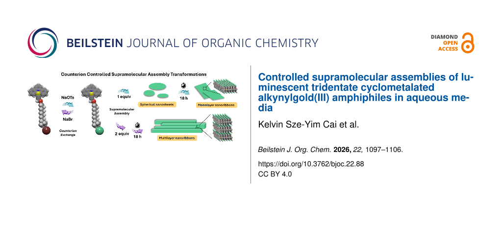
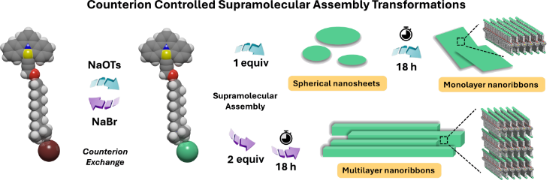
Gold(III) complexes and amphiphiles have been extensively investigated over a decade. Supramolecular assemblies of gold(III) amphiphiles in aqueous media exhibit high sensitivities to external stimulations for soft functional materials. Herein, we introduce a new molecular design of a tridentate cyclometalated gold(III) amphiphile (GA) with flexible molecular structure modifications. Counterion exchange with sodium tosylate induces notable luminescent enhancement and enables control over supramolecular assembly processes. This approach drives a supramolecular assembly transformation of GA from disordered nanosheets to well-ordered nanoribbons upon the additions of multiple equivalents of counterion, enabling a tunable pathway for controlled supramolecular transformation.
Graphical Abstract
Introduction
Supramolecular assemblies in natural systems, such as lipid bilayers and protein complexes, play essential roles in maintaining proper biological functions [1-3]. These natural structures have inspired developments of artificial supramolecular systems that offer structural diversity through delicate designed non-covalent interactions [4-7]. Amphiphilic molecules combine hydrophilic and hydrophobic groups and the intrinsic dual nature enables these molecules to spontaneously assemble into well-defined nanostructures, such as micelles, vesicles, and nanotubes [7-9]. Through implementations of various stimuli-responsive molecular motifs, amphiphiles can respond to external stimuli, such as light, pH, and heat [4,6,10-33], allowing them to be used in biomedical applications, such as, drug delivery and cancer immunotherapy [34,35]. An alternative molecular design approach is to incorporate organometallic complexes into amphiphile designs, thereby combining the advantageous functional properties of inorganic centers with the self-assembled structural properties of amphiphiles [36-40].
Gold(III) complexes have found wide applications for catalysis [41-44], optoelectronic materials [45,46], and bioconjugation methodologies [47-49], considering their unique luminescence properties and excellent aqueous stability. Amphiphilic gold(III) complexes were firstly reported by Che in 2016, who introduced polyethylene glycol (PEG)-functionalized gold(III) complexes to improve the biocompatibility and aqueous solubility [50]. Later, Che and co-workers developed charged cyclometalated gold(III) complexes, achieving kinetically controlled self-assembly in mixed solvents of acetonitrile and water through variation of counterions and its concentration [51]. Yam and co-workers have reported multiresponsive luminescent cationic gold(III) amphiphiles undergoing the self-assembly process in mixed organic solvents [52]. These reports demonstrate remarkable structural sensitivities to gold(III) amphiphiles molecular designs and the resulting nanostructural changes. In general, gold(III) complexes suffered from low luminescent properties due to the intrinsic low-energy d–d ligand field states and the enriched electrophilicity of the gold(III) metal center. The non-emissive low-lying d–d state of gold(III) complexes quenches the luminescent excited state through thermal equilibrium and energy transfer [53]. Cyclometalated gold(III) complexes incorporated with strong σ-donating alkynyl ligands, reported by Yam, exhibit enhanced luminescent properties under ambient conditions, which is attributed to the strong σ-donating alkynyl ligands [54-58].
Our research team has developed bidentate cyclometalated gold(III) amphiphiles featured with counterion-controlled reversible assembly in aqueous media [59-61]. However, the synthetic complexity and aqueous solubility issues of these molecular designs have hindered the functional applicability towards biomaterials. To overcome these limitations, we herein propose a new class of cyclometalated gold(III) amphiphiles GA featuring a tridentate C^N^C cyclometalated core connected to a strong σ-donating alkynyl ligand bearing a propargyl group linked to a terminal quaternary ammonium group through a nonyl linker. Compared with our earlier bidentate designs, this tridentate design provides greater structural rigidity and enhanced luminescence in aqueous media and ambident conditions. Furthermore, the synthetic pathway in this work offers significantly increased accessibility and flexibility of the molecular structural modifications. The incorporation of the propargyl–nonyl–quaternary ammonium motif not only enhances aqueous solubility but also enables fine adjustments of the packing parameter of GA through counterion exchange, thereby achieving a controllable transformation of supramolecular nanostructures. With meticulous investigations of self-assembly processes, photophysical properties, and counterion-dependent structural transformations of GA, a new molecular framework for counterion-controlled luminescent soft materials can be established towards biomedical and biomaterial applications.
Results and Discussion
Design and synthesis of GA
The gold amphiphile was designed with a tridentate C^N^C cyclometalated gold(III) complex core, which is functionalized with a σ-donating alkynyl ligand. A propargyl group was connected by a nonyl-linker with a quaternary ammonium ion as the charged end group (Scheme 1). The synthetic route of GA is shown in Scheme 2. Compound 1 was obtained by the substitution of propargyl bromide with 9-bromo-1-nonanol. Based on the previously reported procedure, the cyclometalated gold(III) complex 2 was synthesized and further reacted with compound 1 in the presence of triethylamine and a catalytic amount of copper iodide to afford the amphiphile precursor 3. The nucleophilic substitution of compound 3 with trimethylamine enabled to afford GA. The chemical structures of all newly synthesized compounds and GA were unambiguously characterized by 1H , 13C NMR spectroscopy and high-resolution mass spectrometry (HRMS) (Supporting Information File 1, Figures S8–S13).
![[1860-5397-22-88-i1]](/bjoc/content/inline/1860-5397-22-88-i1.png?scale=2.0&max-width=1024&background=FFFFFF)
Scheme 1: Schematic illustration of the design of GA and the corresponding supramolecular assembly transformations.
Scheme 1: Schematic illustration of the design of GA and the corresponding supramolecular assembly transforma...
![[1860-5397-22-88-i2]](/bjoc/content/inline/1860-5397-22-88-i2.svg?scale=2.0&max-width=1024&background=FFFFFF)
Photophysical properties and assembly of GA
A freshly prepared aqueous solution of GA (13.4 mM, 1.0 wt %) was annealed at 50.0 °C for 5 min and cooled to room temperature as the stock solution. The stock solution of GA was subsequently diluted into a range of concentrations from 0.01 to 1.0 mM, in estimating the critical aggregation concentration (CAC) by static light scattering measurements (SLS). The CAC of GA was determined as 10 µM (Supporting Information File 1, Figure S1), representing plausible formation of GA supramolecular assemblies in the aqueous environment. A diluted solution of GA (200 µM) was further thermally annealed by heating to 50.0 °C for 5 min and slow cooling to 20.0 °C at a rate of 1.0 °C/min. The UV–vis absorption spectrum of the solution shows an intense absorption band at 304–318 nm and a moderately intense vibronic-structured absorption band at 360–405 nm at 20.0 °C (Figure 1a, black line). The vibrational progressional spacings of 1300–1310 cm−1 in the lower energy band represent the skeletal vibrational frequencies of the cyclometalated C^N^C ligand. In considering similar absorption bands observed in the cyclometalated gold(III) complex 2 [62] and the related alkynylgold(III) complexes [54-56], the vibronic-structured absorption band at 360–405 nm is revealed as insensitive to the structurally modified alkynyl ligands, which is generally assigned as a metal-perturbed intraligand (IL) π–π* transition of the C^N^C ligand. A subtle increase in absorbance with a hypochromic-shift of 3 nm was observed upon heating the solution to 50.0 °C (Figure 1a, red line), which was reversed upon cooling to 20.0 °C. This thermal annealing process is fully reversible with clear isosbestic points, suggesting that the supramolecular assembly of GA in aqueous media is under thermodynamic control without observable degradation. A methanol solution of GA (200 µM) shows no signs of spectral shift and is reversible upon annealing (Supporting Information File 1, Figure S2a), indicating the absence of supramolecular aggregation in an organic environment and the thermal stability of GA in both organic and aqueous media.
![[1860-5397-22-88-1]](/bjoc/content/figures/1860-5397-22-88-1.png?scale=2.0&max-width=1024&background=FFFFFF)
Figure 1: (a) UV–vis absorption spectra of GA (200 μM) in MQ water heated to 50.0 °C (red line) and cooled to 20.0 °C (black line) at a rate of 1.0 °C/min. (b) Emission spectra of GA (200 μM) in MQ water upon decreasing the temperature from 50.0 °C (black line) to 10.0 °C (purple line).
Figure 1: (a) UV–vis absorption spectra of GA (200 μM) in MQ water heated to 50.0 °C (red line) and cooled to...
Upon excitation a methanol solution of GA (200 µM) with 365 nm, a broad and structureless emission band at 428 nm is observed at 20.0 °C (Supporting Information File 1, Figure S2b, green line). In contrast, an aqueous solution of GA (200 µM) exhibits an additional vibronic-structured emission band at 465–540 nm, and the intensity of the emission band at 429 nm (Figure 1b, green-line) is lower than that of GA in organic media (Supporting Information File 1, Figure S2b, green line) at 20.0 °C. Given that a dichloromethane solution of cyclometalated gold(III) complex 2 (200 µM) shows significantly lowered emission intensity at 430 nm (Supporting Information File 1, Figure S3), the emission spectral results suggested that the luminescent properties of GA have been greatly improved through the coupling with a σ-donor to gold(III) complex. Upon progressively heating the aqueous solution of GA from 10.0 °C to 50.0 °C, the resulting emission intensity of the vibronic-structured emission band was gradually decreased along with the subtly decreased structureless emission band at 428 nm (Figure 1b), possibly due to emissions from the GA assemblies.
Upon increasing the water content in methanol, the UV–vis absorption spectra of GA show a progressive increase in absorbance of the IL band tail at 420 nm (Supporting Information File 1, Figure S4a and S4b), suggesting the formation of aggregates. The emission spectra of GA show a reduction in intensity of the structureless emission band at 429 nm with increased emission of the lower-energy vibronic-structured emission band at 465–540 nm upon water content increased from 0% to 99% (Supporting Information File 1, Figure S4c and S4d). The obtained results demonstrated a clear redistribution of emission intensity due to aggregation of GA in aqueous media. The rigid molecular packed structures of GA in the aggregated form restrict intramolecular vibrations of the C^N^C ligand and suppress non-radiative decay pathways to activate the lower-energy emission. An essentially identical emission spectrum was observed for the methanol solution of GA at 20.0 °C (Supporting Information File 1, Figure S2b, green line) to that of an aqueous solution of GA at 50 °C (Figure 1b, black line). This can be illustrated by the vibronic-structured emission band quenched at increased temperature and in the organic medium due to possible non-radiative decay pathways. These results further confirmed that the vibronic-structure emission band can be induced upon GA assembly, which shows possible correlations with temperature and water content.
Counterion-controlled supramolecular assembly transformations of GA
Negative-stained transmission electron microscopy (TEM) was used to analyze the assembled nanostructures of GA. TEM images of a thermally annealed aqueous solution of GA (2.68 mM, 0.2 wt %) reveal sheet-like nanostructures with widths ranging from hundreds of nanometers to micrometers (Figure 2a). To a thermally annealed aqueous solution of GA (200 μM) were added varying equivalents of an aqueous solution of sodium tosylate (1.0, 2.0, and 4.0 equiv), and subsequently heated to 50.0 °C and slowly cooled to 20.0 °C at a rate of 1.0 °C/min. The UV–vis absorption spectra of the solution show a progressive bathochromic shift with increasing sodium tosylate equivalents (Figure 2b). The presence of isosbestic points at 359 nm, 375 nm, 381 nm, 395 nm, and 401 nm within the vibronic-structured absorption band indicates a selective counterion exchange process, where the organic tosylate anion replaces the inorganic bromide anion. The corresponding emission spectra of the solution show a notable enhancement in the overall intensity upon the addition of 1.0–2.0 equiv of sodium tosylate (Figure 2c red line and blue line). However, the vibronic-structured emission band diminished upon the addition of 4.0 equiv of sodium tosylate (Figure 2c, green line).
![[1860-5397-22-88-2]](/bjoc/content/figures/1860-5397-22-88-2.png?scale=2.0&max-width=1024&background=FFFFFF)
Figure 2: (a) TEM image of a thermally annealed aqueous solution of GA (2.68 mM). (b) UV–vis absorption spectra and (c) emission spectra of GA (200 μM) in Milli-Q water at 20.0 °C after addition of sodium tosylate (1.0, 2.0, and 4.0 equiv).
Figure 2: (a) TEM image of a thermally annealed aqueous solution of GA (2.68 mM). (b) UV–vis absorption spect...
The time-dependent UV–vis adsorption spectra of the annealed solution of GA without the addition of sodium tosylate show no significant changes after 18 h at 20.0 °C (Supporting Information File 1, Figure S5a). In contrast, the absorbance of the annealed solution of GA with 1.0, 2.0 and 4.0 equiv of sodium tosylate gradually increased after 18 hours of aging (Figure 3a, and Figures S5b and S5c in Supporting Information File 1), indicating the formation of a thermodynamically driven assembly. A clear lag time for GA with tosylate (1.0 equiv) suggests the presence of a metastable assembly state (Figure 3b, red line). The spontaneous assembly process is suppressed due to stronger intermolecular interactions between the GA cation end group and the tosylate anion than with the bromide anion [51,63]. Increasing the concentration of sodium tosylate from 1.0 to 4.0 equiv progressively reduces the lag time and accelerates the assembly process (Figure 3b). Notably, the final absorbance intensity of GA with tosylate (2.0 and 4.0 equiv) is approximately twice that of GA with tosylate (1.0 equiv), suggesting a significant difference in the density or size of the thermodynamically driven assemblies formed (Figure 3b).
![[1860-5397-22-88-3]](/bjoc/content/figures/1860-5397-22-88-3.png?scale=2.0&max-width=1024&background=FFFFFF)
Figure 3: (a) Time-dependent UV–vis adsorption spectra of GA (200 μM) with addition of 1.0 equiv of sodium tosylate in Milli-Q water at 20.0 °C. (b) Time-dependent absorbance changes of GA at 420 nm with and without the addition of sodium tosylate (1, 2, or 4 equiv).
Figure 3: (a) Time-dependent UV–vis adsorption spectra of GA (200 μM) with addition of 1.0 equiv of sodium to...
A freshly prepared thermally annealed aqueous solution of GA (2.68 mM, 0.2 wt %) with tosylate (1.0 equiv) was imaged with spherical nanosheets with diameters ranging from ≈200 nm to 2 µm (Figure 4a). After being aged for 18 h, the spherical nanosheets of GA have evolved into stacked nanoribbons (Figure 4b). The time-dependent transitions indicate a kinetically controlled assembly process, in which the initial metastable assemblies transform into their thermodynamically driven assemblies. Upon addition of 2.0 equiv of tosylate and aging for 18 h, long nanoribbons were observed (Figure 4c). In contrast, irregular assemblies were observed in an aged GA solution with 4.0 equiv of tosylate (Supporting Information File 1, Figure S6), possibly due to the disruption of ordered assemblies by supersaturated ions. Atomic force microscopy (AFM) was performed to further confirm the assembled nanostructure of the aged GA solution (2.68 mM, 0.2 wt %) with tosylate (1.0 and 2.0 equiv). Essentially identical nanoribbon structures were found with a uniform height of 3.8 nm and 25 nm (Figure 4d and 4e). Given that the molecular length of GA is estimated to be approximately 2.4 nm, it is assumed that the nanoribbons of GA with 1.0 equiv of tosylate are monolayers in which two GA molecules exhibit head-to-tail stacking, while GA with 2.0 equiv of tosylate forms multilayer nanoribbons. These results demonstrate that counterion exchange from bromide to tosylate enhances the packing parameter of GA, resulting in a transition from less ordered nanosheets to well-ordered nanoribbons. In considering the significant influence of counterions, to the solution of GA (2.68 mM, 0.2 wt %) with tosylate (1.0 and 2.0 equiv) were added the corresponding equivalents of sodium bromide followed by further thermal annealing. The TEM images of the resulting solution revealed the reverse supramolecular transformation from regular nanoribbons to sheet-like nanostructures (Supporting Information File 1, Figure S7).
![[1860-5397-22-88-4]](/bjoc/content/figures/1860-5397-22-88-4.png?scale=2.0&max-width=1024&background=FFFFFF)
Figure 4: Time-dependent TEM changes of the thermally annealed GA solution (2.68 mM) with the addition of 1.0 equiv of sodium tosylate (a) freshly prepared (inset: higher magnification of spherical nanosheets, scale bar: 200 nm) and (b) after 18 h. (c) TEM image of the thermally annealed GA solution (2.68 mM) with the addition of 2.0 equiv of sodium tosylate obtained at 18 h (inset: higher magnification of long nanoribbons, scale bar: 500 nm). AFM images of the aged GA solution (2.68 mM) with the addition of (d) 1.0 equiv and (e) 2.0 equiv of sodium tosylate (inset: height profile along the indicated line).
Figure 4: Time-dependent TEM changes of the thermally annealed GA solution (2.68 mM) with the addition of 1.0...
The distinct assembly outcomes observed with bromide and tosylate can be rationalized by considering their effects on the molecular packing parameter. As a small, highly hydrated inorganic anion, bromide maintains a larger effective head group area, resulting in a packing parameter that favours the formation of relatively flat but disordered lamellar nanosheets. In contrast, tosylate, being a larger and more hydrophobic organic anion with an aromatic moiety, reduces the effective head group area through lower hydration and additional π–π stacking and hydrophobic interactions. This shift in the packing parameter promotes anisotropic growth, enabling the transformation from disordered nanosheets into well-ordered monolayer nanoribbons and eventually multilayer nanoribbons with increasing tosylate concentration. These findings highlight the critical role of counterion nature in directing the hierarchical organization of gold(III) amphiphiles in aqueous media.
Conclusion
A tridentate cyclometalated gold(III) amphiphile, functionalized with a σ-donating alkynyl ligand, was designed and synthesized. This structural modification significantly enhanced its luminescent properties in aqueous media. Supramolecular assembled nanostructures of GA were imaged and confirmed by TEM and AFM. The addition of sodium tosylate, enabled a thermodynamically driven assembly process, leading to supramolecular assembly transformations from sheet-like nanostructures to spherical nanosheets, and to stacked nanoribbons throughout an aging process. Well-defined monolayered and multilayered nanoribbons were observed by varying concentrations of tosylate ions. The reverse supramolecular transformation was enabled in the current gold(III) amphiphilic design by the addition of bromide ions. The current system of a tridentate cyclometalated gold(III) amphiphile lays the foundation for developing next generations of controlled supramolecular assembly of gold(III) amphiphiles.
Experimental
Materials: All commercial reagents were purchased from Acros Organics, Aladdin, Bidepharm, Dieckmann, Macklin and Tokyo Chemical Industry Co. Ltd, and were used as received unless otherwise specified. All reactions were performed under nitrogen unless otherwise specified. Analytical thin-layer chromatography (TLC) was performed with Macherey-Nagel Silica gel 60 UV254 aluminum plates and visualization was accomplished by UV light (254 nm) or staining with phosphomolybdic acid (PMA) followed by heating. Flash column chromatography was performed using Macherey-Nagel Silica gel 60 (230–400 mesh). Deuterated solvents were purchased from Cambridge Isotope Laboratories Inc.
Compound 1: Synthesis according to the modification of a reported procedure [64]. A mixture of 9-bromo-1-nonanol (482 mg, 2.16 mmol) and sodium hydride (100 mg, 4.17 mmol) in dimethylformamide (5 mL) was stirred at 0 °C for 15 min. Propargyl bromide (0.2 mL, 2.59 mmol) was added dropwise to the mixture solution and stirred at room temperature for 16 h. The resulting mixture was washed with brine (25 mL × 1), water (25 mL × 2), and brine (25 mL × 1). The organic layer was dried over sodium sulfate. The solvent was evaporated under vacuum and the residue subjected to column chromatography on silica gel (hexane/ethyl acetate 10:1 (v/v, Rf = 0.5) to afford compound 1 as pale-yellow liquid (284 mg, 1.09 mmol) in 50% yield. 1H NMR (600 MHz, CDCl3) δ 4.08 (s, 2H), 3.46 (t, J = 6.6 Hz, 2H), 3.36 (t, J = 7.0 Hz, 2H), 2.39 (s, 1H), 1.80 (q, J = 7.3 Hz, 2H), 1.54 (t, J = 6.4 Hz, 2H), 1.37 (t, J = 7.4 Hz, 2H), 1.31–1.25 (m, 8H); HRMS–ESI+ (m/z): [M + H]+ calcd for C12H22BrO, 261.0849; found, 261.0848.
Compound 2: Synthesis according to the modification of a reported procedure [62]. 1H NMR (600 MHz, CD3SOCD3) δ 8.19 (t, J = 8.0 Hz, 1H), 8.01–7.97 (m, 2H), 7.89 (d, J = 7.7 Hz, 2H), 7.70 (d, J = 7.2 Hz, 2H), 7.43 (t, J = 7.3 Hz, 2H), 7.32 (t, J = 7.6 Hz, 2H).
Compound 3: A mixture of compound 1 (87 mg, 0.33 mmol) and compound 2 (103 mg, 0.22 mmol) in the presence of a catalytic amount of copper(I) iodide (4 mg, 0.02 mmol) in triethylamine (1 mL) and dichloromethane (20 mL) was stirred at room temperature for 3 h. The resulting mixture was dried under vacuum and subjected to column chromatography on silica gel (hexane/dichloromethane 1:2 (v/v, Rf = 0.5) to afford compound 3 as yellow solid (109 mg, 0.16 mmol) in 71% yield. 1H NMR (400 MHz, CDCl3) δ 7.98 (d, J = 7.3 Hz, 2H), 7.83 (t, J = 7.9 Hz, 1H), 7.51 (d, J = 7.7 Hz, 2H), 7.43 (d, J = 7.9 Hz, 2H), 7.35 (t, J = 6.7 Hz, 2H), 7.22 (t, J = 7.6 Hz, 2H), 4.47 (s, 2H), 3.73 (t, J = 6.7 Hz, 2H), 3.38 (t, J = 6.9 Hz, 2H), 1.83 (p, J = 7.0 Hz, 2H), 1.76–1.64 (m, 2H), 1.42 (dt, J = 14.4, 7.6 Hz, 4H), 1.32 (q, J = 5.6 Hz, 6H); 13C NMR (101 MHz, CDCl3) δ 166.85, 165.10, 149.11, 142.35, 136.77, 132.06, 126.85, 125.27, 116.85, 96.68, 86.21, 77.36, 69.56, 60.35, 34.25, 33.00, 29.88, 29.59, 28.89, 28.34, 26.46; HRMS–ESI+ (m/z): [M + H]+ calcd for C29H32AuBrNO, 686.1328; found, 686.1319.
GA: A mixture of compound 3 (92 mg, 0.13 mmol) and 20% trimethylamine in ethanol (1 mL) in dichloromethane (2 mL) was heated at 50 °C for 24 h in a Schlenk tube. The resulting mixture was cooled to room temperature and precipitated with dichloromethane and diethyl ether. The precipitate was filtered off and was washed with hexane to afford GA as pale-yellow solid (69 mg, 0.09 mmol) in 69% yield. 1H NMR (600 MHz, MeOD) δ 8.02–7.97 (m, 1H), 7.86 (d, J = 7.7 Hz, 2H), 7.70 (t, J = 7.9 Hz, 4H), 7.31 (t, J = 6.6 Hz, 2H), 7.27–7.21 (m, 2H), 4.40 (s, 2H), 3.73 (t, J = 6.3 Hz, 2H), 3.26 (s, 2H), 3.08 (s, 9H), 1.76 (q, J = 8.0 Hz, 2H), 1.69 (q, J = 7.1 Hz, 2H), 1.48 (d, J = 7.1 Hz, 2H), 1.38 (d, J = 25.2 Hz, 8H); 13C NMR (151 MHz, MeOD) δ 167.70, 166.23, 150.82, 144.43, 137.36, 132.60, 128.01, 126.67, 118.53, 70.40, 60.99, 53.43, 30.61, 30.40, 30.28, 30.06, 27.29, 23.88; HRMS–ESI+ (m/z): [M – Br]+ calcd for C32H40AuN2O, 665.2801; found, 665.2846.
Preparation of aqueous sample: GA (13.4 mM, 1.0 wt %) was dissolved in fresh Milli-Q water (MQ water). The solution was heated at 50.0 °C for 5 min, then slowly cooled to 20.0 °C at a rate of 1.0 °C/min to form assembled structures.
UV–vis spectroscopy: UV–vis measurements were performed on an Agilent Cary 60 UV–vis spectrophotometer with a 1 cm path length quartz cuvette. A Luma 40/8453 temperature-controlled cuvette holder with four optical ports was mounted in the sample compartment of the Agilent Cary 60 UV–vis spectrophotometer. Time-dependent UV–vis adsorption measurements were performed using a small stirring bar (3 × 5 mm) at a constant stirring speed of 1200 rpm. Slits were installed on the cuvette holder to prevent the stirring bar from interfering with the optical path to the spectrometer. Measurements of all samples were carried out at 20.0 °C unless otherwise specified.
Fluorescence spectroscopy: Fluorescence measurements were performed on an Agilent G9800A Cary Eclipse fluorescence spectrophotometer with a 1 cm path length quartz cuvette. A Luma 40/8453 temperature-controlled cuvette holder with four optical ports was mounted in the sample compartment of the Agilent G9800A Cary Eclipse fluorescence spectrophotometer. Measurements of all samples were carried out at 20.0 °C unless otherwise specified.
Static light scattering (SLS): Static light scattering intensities of each sample were determined on a Wyatt Technology DynaPro NanoStar. The scattering intensities were recorded as a parameter for assembly size, given that the objects in solution are anisotropic and the models used by Wyatt software are fitting for spherical objects. The critical aggregation concentration (CAC) of GA was determined by the scattering intensities of the solutions of GA (concentration: 0.01 to 1.0 mM) at 20.0 °C. The scattering rate was normalized by the concentration of the solution to yield the molar scattering intensity (M counts s−1 M−1). Ten replications were performed, and the data was averaged to show the molar scattering intensity and corresponding standard deviation.
Treatment of GA with sodium tosylate for counter anion-controlled supramolecular transformation: To a thermally annealed aqueous solution of GA (13.4 mM) was added an aqueous solution of sodium tosylate (1.0, 2.0, and 4.0 equiv). The obtained solution was heated at 50.0 °C for 5 min, and slowly cooled to 20.0 °C at a rate of 1.0 °C/min.
Transmission electron microscopy (TEM): TEM samples were prepared by depositing sample solutions (0.2 wt %, 10.0 μL) onto a carbon grid (Micro to Nano, EMR Carbon support film on copper, 400 square mesh) for 30 s. The sample solution was removed by blotting and UranyLess EM stain solution (Electron Microscopy Science, 5.0 μL) was directly deposited onto the grid for 15 s, and the stain was removed by blotting. Grids were observed in a ThermoFisher Talos L120C transmission electron microscope at an accelerating voltage of 120 kV. TEM images were captured on a Ceta 16M CCD camera.
Atomic force microscopy (AFM): The atomic force microscopy was performed on a JPK NanoWizard V BioScience AFM (Bruker) equipped on Nikon ECLIPSE Ti-2 microscope with a cantilever (RFESPG-75, Bruker). The measurement of the assembled morphology was conducted in air with the Peakforce Tapping mode. The sample solution (0.2 wt %, 5.0 μL) was dropped onto a freshly cleaved mica plate surface and spin-coated to remove excess solvent.
Supporting Information
| Supporting Information File 1: Experimental details, supporting figures, and copies of spectra. | ||
| Format: PDF | Size: 1.5 MB | Download |
Data Availability Statement
All data that supports the findings of this study is available in the published article and/or the supporting information of this article.
References
-
Simmons, N. S.; Blout, E. R. Biophys. J. 1960, 1, 55–62. doi:10.1016/s0006-3495(60)86875-0
Return to citation in text: [1] -
Lino, T. J. Supramol. Struct. 1974, 2, 372–384. doi:10.1002/jss.400020226
Return to citation in text: [1] -
Chapman, D. Q. Rev. Biophys. 1975, 8, 185–235. doi:10.1017/s0033583500001797
Return to citation in text: [1] -
Aida, T.; Meijer, E. W.; Stupp, S. I. Science 2012, 335, 813–817. doi:10.1126/science.1205962
Return to citation in text: [1] [2] -
Krieg, E.; Bastings, M. M. C.; Besenius, P.; Rybtchinski, B. Chem. Rev. 2016, 116, 2414–2477. doi:10.1021/acs.chemrev.5b00369
Return to citation in text: [1] -
Feringa, B. L. Angew. Chem., Int. Ed. 2017, 56, 11060–11078. doi:10.1002/anie.201702979
Return to citation in text: [1] [2] -
Chen, S.; Costil, R.; Leung, F. K.-C.; Feringa, B. L. Angew. Chem., Int. Ed. 2021, 60, 11604–11627. doi:10.1002/anie.202007693
Return to citation in text: [1] [2] -
Israelachvili, J. N.; Mitchell, D. J.; Ninham, B. W. J. Chem. Soc., Faraday Trans. 2 1976, 72, 1525. doi:10.1039/f29767201525
Return to citation in text: [1] -
Degiorgio, V. Europhys. News 1985, 16, 9–12. doi:10.1051/epn/19851606009
Return to citation in text: [1] -
Shigemitsu, H.; Fujisaku, T.; Tanaka, W.; Kubota, R.; Minami, S.; Urayama, K.; Hamachi, I. Nat. Nanotechnol. 2018, 13, 165–172. doi:10.1038/s41565-017-0026-6
Return to citation in text: [1] -
Dattler, D.; Fuks, G.; Heiser, J.; Moulin, E.; Perrot, A.; Yao, X.; Giuseppone, N. Chem. Rev. 2020, 120, 310–433. doi:10.1021/acs.chemrev.9b00288
Return to citation in text: [1] -
Baroncini, M.; Silvi, S.; Credi, A. Chem. Rev. 2020, 120, 200–268. doi:10.1021/acs.chemrev.9b00291
Return to citation in text: [1] -
Volarić, J.; Szymanski, W.; Simeth, N. A.; Feringa, B. L. Chem. Soc. Rev. 2021, 50, 12377–12449. doi:10.1039/d0cs00547a
Return to citation in text: [1] -
Leung, F. K.-C. Aqueous Supramolecular Assemblies of Photocontrolled Molecular Amphiphiles. In Supramolecular Assemblies Based on Electrostatic Interactions; Aboudzadeh, M. A.; Frontera, A., Eds.; Springer International Publishing: Cham, Switzerland, 2022; pp 267–308. doi:10.1007/978-3-031-00657-9_9
Return to citation in text: [1] -
Cheung, L.-H.; Liu, B. B.; Leung, F. K.-C. Polym. J. 2023, 55, 1189–1198. doi:10.1038/s41428-023-00792-7
Return to citation in text: [1] -
Chau, A. K.-H.; Leung, F. K.-C. Adv. Colloid Interface Sci. 2023, 315, 102892. doi:10.1016/j.cis.2023.102892
Return to citation in text: [1] -
Cheung, L.-H.; To, J. C.; Wong, W.-K.; Stuart, M. C. A.; Kajitani, T.; Keng, V. W.; Leung, F. K.-C. ACS Appl. Mater. Interfaces 2024, 16, 4056–4070. doi:10.1021/acsami.3c16795
Return to citation in text: [1] -
Hung, K.-L.; Cheung, L.-H.; Ren, Y.; Chau, M.-H.; Lam, Y.-Y.; Kajitani, T.; Leung, F. K.-C. Beilstein J. Org. Chem. 2024, 20, 1590–1603. doi:10.3762/bjoc.20.142
Return to citation in text: [1] -
Yau, J. C.-K.; Hung, K.-L.; Ren, Y.; Kajitani, T.; Stuart, M. C. A.; Leung, F. K.-C. J. Colloid Interface Sci. 2024, 662, 391–403. doi:10.1016/j.jcis.2024.02.075
Return to citation in text: [1] -
Meng, J.; Cheung, L.-H.; Ren, Y.; Stuart, M. C. A.; Wang, Q.; Chen, S.; Chen, J.; Leung, F. K.-C. Macromol. Rapid Commun. 2024, 45, 2400261. doi:10.1002/marc.202400261
Return to citation in text: [1] -
Chau, M.-H.; Wong, W.-K.; Kajitani, T.; Leung, F. K.-C. Adv. Sci. 2024, 11, 2407130. doi:10.1002/advs.202407130
Return to citation in text: [1] -
Chau, M.-H.; Wong, W.-K.; Meng, S.; Kajitani, T.; Leung, F. K.-C. Dyes Pigm. 2024, 230, 112334. doi:10.1016/j.dyepig.2024.112334
Return to citation in text: [1] -
Wycisk, V.; Wagner, M.-C.; Urner, L. H. ChemPlusChem 2024, 89, e202300594. doi:10.1002/cplu.202300594
Return to citation in text: [1] -
Hung, K.-L.; Wong, W.-K.; Chau, M.-H.; Yau, J. C.-K.; Kajitani, T.; Chen, S.; Leung, F. K.-C. ACS Appl. Nano Mater. 2025, 8, 8876–8885. doi:10.1021/acsanm.5c00964
Return to citation in text: [1] -
Cheung, L.-H.; Leung, F. K.-C. Chem. – Eur. J. 2025, 31, e202500791. doi:10.1002/chem.202500791
Return to citation in text: [1] -
Yau, J. C.-K.; Leung, F. K.-C. J. Organomet. Chem. 2025, 1031, 123596. doi:10.1016/j.jorganchem.2025.123596
Return to citation in text: [1] -
Liu, B. B.; Zhang, H.; Wong, W.-K.; Chau, M.-H.; Cheung, L.-H.; Kajitani, T.; Leung, F. K.-C. Small 2025, 21, 2503359. doi:10.1002/smll.202503359
Return to citation in text: [1] -
Meng, S.; Chau, M.-H.; Kajitani, T.; Leung, F. K.-C. Adv. Rob. Res. 2026, 2, e202500070. doi:10.1002/adrr.202500070
Return to citation in text: [1] -
Yau, J. C.-K.; Chen, C. Y.; Zhang, H.; Chau, M.-H.; Kajitani, T.; Leung, F. K.-C. Mater. Chem. Front. 2025, 9, 3414–3424. doi:10.1039/d5qm00491h
Return to citation in text: [1] -
Ren, Y.; Cheung, L.-H.; Kajitani, T.; Leung, F. K.-C. J. Colloid Interface Sci. 2026, 702, 138850. doi:10.1016/j.jcis.2025.138850
Return to citation in text: [1] -
Lee, T. H.-W.; Hung, K.-L.; Kajitani, T.; Leung, F. K.-C. ChemPhotoChem 2026, 10, e202500333. doi:10.1002/cptc.202500333
Return to citation in text: [1] -
Zhang, H.; Cai, K. S.-Y.; Liu, B. B.; Huang, J.; Leung, F. K.-C. Langmuir 2026, 42, 10337–10347. doi:10.1021/acs.langmuir.5c06496
Return to citation in text: [1] -
Leung, F.; Hung, K.-L.; Wong, W. K. R.; Cheung, L.-H.; Chau, M.-H.; Kajitani, T. Res. Square 2025. doi:10.21203/rs.3.rs-7996724/v1
Return to citation in text: [1] -
Yin, B.; Wong, W.-K.; Ng, Y.-M.; Yang, M.; Leung, F. K.-C.; Wong, D. S.-H. Pharmaceutics 2023, 15, 1427. doi:10.3390/pharmaceutics15051427
Return to citation in text: [1] -
Wong, W.-K.; Ren, Y.; Leung, F. K.-C. Photothermal-chemotherapy: the emerging supramolecular photothermal molecules and the recent advances. In Nanophototherapy; Manivasagan, P.; Venkatesan, J.; Jang, E.-S., Eds.; Elsevier: Amsterdam, Netherlands, 2025; pp 463–499. doi:10.1016/b978-0-443-13937-6.00007-x
Return to citation in text: [1] -
Owen, T.; Butler, A. Coord. Chem. Rev. 2011, 255, 678–687. doi:10.1016/j.ccr.2010.12.009
Return to citation in text: [1] -
Mayoral Muñoz, M. J.; Fernández, G. Chem. Sci. 2012, 3, 1395–1398. doi:10.1039/c2sc01101h
Return to citation in text: [1] -
Fu, H. L.-K.; Yam, V. W.-W. Chem. Lett. 2018, 47, 605–610. doi:10.1246/cl.180096
Return to citation in text: [1] -
Yam, V. W.-W.; Law, A. S.-Y. J. Chin. Chem. Soc. 2020, 67, 2246–2252. doi:10.1002/jccs.202000536
Return to citation in text: [1] -
Poon, J. K.-L.; Chen, Z.; Leung, S. Y.-L.; Leung, M.-Y.; Yam, V. W.-W. Proc. Natl. Acad. Sci. U. S. A. 2021, 118, e2022829118. doi:10.1073/pnas.2022829118
Return to citation in text: [1] -
Zi, W.; Toste, F. D. Chem. Soc. Rev. 2016, 45, 4567–4589. doi:10.1039/c5cs00929d
Return to citation in text: [1] -
Cui, J.-F.; Ko, H.-M.; Shing, K.-P.; Deng, J.-R.; Lai, N. C.-H.; Wong, M.-K. Angew. Chem., Int. Ed. 2017, 129, 3074–3079. doi:10.1002/anie.201612243
Return to citation in text: [1] -
Rocchigiani, L.; Bochmann, M. Chem. Rev. 2021, 121, 8364–8451. doi:10.1021/acs.chemrev.0c00552
Return to citation in text: [1] -
Kumar, R.; Nevado, C. Angew. Chem., Int. Ed. 2017, 56, 1994–2015. doi:10.1002/anie.201607225
Return to citation in text: [1] -
Yam, V. W.-W.; Chan, A. K.-W.; Hong, E. Y.-H. Nat. Rev. Chem. 2020, 4, 528–541. doi:10.1038/s41570-020-0199-7
Return to citation in text: [1] -
Tang, M.-C.; Chan, M.-Y.; Yam, V. W.-W. Chem. Rev. 2021, 121, 7249–7279. doi:10.1021/acs.chemrev.0c00936
Return to citation in text: [1] -
Kung, K. K.-Y.; Ko, H.-M.; Cui, J.-F.; Chong, H.-C.; Leung, Y.-C.; Wong, M.-K. Chem. Commun. 2014, 50, 11899–11902. doi:10.1039/c4cc04467c
Return to citation in text: [1] -
Sit, H.-Y.; Yang, B.; Ka‐Yan Kung, K.; Siu‐Lun Tam, J.; Wong, M.-K. ChemPlusChem 2019, 84, 1739–1743. doi:10.1002/cplu.201900612
Return to citation in text: [1] -
Ko, H.-M.; Deng, J.-R.; Cui, J.-F.; Kung, K. K.-Y.; Leung, Y.-C.; Wong, M.-K. Bioorg. Med. Chem. 2020, 28, 115375. doi:10.1016/j.bmc.2020.115375
Return to citation in text: [1] -
Wang, F.; Lan, M.; To, W.-P.; Li, K.; Lok, C.-N.; Wang, P.; Che, C.-M. Chem. Commun. 2016, 52, 13273–13276. doi:10.1039/c6cc06767k
Return to citation in text: [1] -
Wan, Q.; Xia, J.; Lu, W.; Yang, J.; Che, C.-M. J. Am. Chem. Soc. 2019, 141, 11572–11582. doi:10.1021/jacs.9b04007
Return to citation in text: [1] [2] -
Leung, M.-Y.; Leung, S. Y.-L.; Yim, K.-C.; Chan, A. K.-W.; Ng, M.; Yam, V. W.-W. J. Am. Chem. Soc. 2019, 141, 19466–19478. doi:10.1021/jacs.9b10607
Return to citation in text: [1] -
Yam, V. W.-W.; Choi, S. W.-K.; Lai, T.-F.; Lee, W.-K. J. Chem. Soc., Dalton Trans. 1993, 1001. doi:10.1039/dt9930001001
Return to citation in text: [1] -
Yam, V. W.-W.; Wong, K. M.-C.; Hung, L.-L.; Zhu, N. Angew. Chem., Int. Ed. 2005, 44, 3107–3110. doi:10.1002/anie.200500253
Return to citation in text: [1] [2] -
Wong, K. M.-C.; Zhu, X.; Hung, L.-L.; Zhu, N.; Yam, V. W.-W.; Kwok, H.-S. Chem. Commun. 2005, 2906–2908. doi:10.1039/b503315b
Return to citation in text: [1] [2] -
Wong, K. M.-C.; Hung, L.-L.; Lam, W. H.; Zhu, N.; Yam, V. W.-W. J. Am. Chem. Soc. 2007, 129, 4350–4365. doi:10.1021/ja068264u
Return to citation in text: [1] [2] -
Au, V. K.-M.; Wong, K. M.-C.; Zhu, N.; Yam, V. W.-W. J. Am. Chem. Soc. 2009, 131, 9076–9085. doi:10.1021/ja9027692
Return to citation in text: [1] -
Au, V. K.-M.; Wong, K. M.-C.; Tsang, D. P.-K.; Chan, M.-Y.; Zhu, N.; Yam, V. W.-W. J. Am. Chem. Soc. 2010, 132, 14273–14278. doi:10.1021/ja106579d
Return to citation in text: [1] -
Jiang, J.-J.; Chau, A. K.-H.; Wong, M.-K.; Leung, F. K.-C. Eur. J. Inorg. Chem. 2022, e202200281. doi:10.1002/ejic.202200281
Return to citation in text: [1] -
Chau, M.-H.; Chan, A. K.-H.; Ren, Y.; Jiang, J.-J.; Wong, M.-K.; Leung, F. K.-C. J. Organomet. Chem. 2023, 999, 122827. doi:10.1016/j.jorganchem.2023.122827
Return to citation in text: [1] -
Chan, A. K.-H.; Chau, M.-H.; Ren, Y.; Jiang, J.-J.; Wong, M.-K.; Leung, F. K.-C. ChemPlusChem 2024, 89, e202300316. doi:10.1002/cplu.202300316
Return to citation in text: [1] -
Wong, K.-H.; Cheung, K.-K.; Chan, M. C.-W.; Che, C.-M. Organometallics 1998, 17, 3505–3511. doi:10.1021/om980274j
Return to citation in text: [1] [2] -
Liu; González, Y. I.; Danino, D.; Kaler, E. W. Macromolecules 2005, 38, 2482–2491. doi:10.1021/ma047646m
Return to citation in text: [1] -
Chen, J.; Wang, J.; Bai, Y.; Li, K.; Garcia, E. S.; Ferguson, A. L.; Zimmerman, S. C. J. Am. Chem. Soc. 2018, 140, 13695–13702. doi:10.1021/jacs.8b06875
Return to citation in text: [1]
| 64. | Chen, J.; Wang, J.; Bai, Y.; Li, K.; Garcia, E. S.; Ferguson, A. L.; Zimmerman, S. C. J. Am. Chem. Soc. 2018, 140, 13695–13702. doi:10.1021/jacs.8b06875 |
| 54. | Yam, V. W.-W.; Wong, K. M.-C.; Hung, L.-L.; Zhu, N. Angew. Chem., Int. Ed. 2005, 44, 3107–3110. doi:10.1002/anie.200500253 |
| 55. | Wong, K. M.-C.; Zhu, X.; Hung, L.-L.; Zhu, N.; Yam, V. W.-W.; Kwok, H.-S. Chem. Commun. 2005, 2906–2908. doi:10.1039/b503315b |
| 56. | Wong, K. M.-C.; Hung, L.-L.; Lam, W. H.; Zhu, N.; Yam, V. W.-W. J. Am. Chem. Soc. 2007, 129, 4350–4365. doi:10.1021/ja068264u |
| 51. | Wan, Q.; Xia, J.; Lu, W.; Yang, J.; Che, C.-M. J. Am. Chem. Soc. 2019, 141, 11572–11582. doi:10.1021/jacs.9b04007 |
| 63. | Liu; González, Y. I.; Danino, D.; Kaler, E. W. Macromolecules 2005, 38, 2482–2491. doi:10.1021/ma047646m |
| 1. | Simmons, N. S.; Blout, E. R. Biophys. J. 1960, 1, 55–62. doi:10.1016/s0006-3495(60)86875-0 |
| 2. | Lino, T. J. Supramol. Struct. 1974, 2, 372–384. doi:10.1002/jss.400020226 |
| 3. | Chapman, D. Q. Rev. Biophys. 1975, 8, 185–235. doi:10.1017/s0033583500001797 |
| 34. | Yin, B.; Wong, W.-K.; Ng, Y.-M.; Yang, M.; Leung, F. K.-C.; Wong, D. S.-H. Pharmaceutics 2023, 15, 1427. doi:10.3390/pharmaceutics15051427 |
| 35. | Wong, W.-K.; Ren, Y.; Leung, F. K.-C. Photothermal-chemotherapy: the emerging supramolecular photothermal molecules and the recent advances. In Nanophototherapy; Manivasagan, P.; Venkatesan, J.; Jang, E.-S., Eds.; Elsevier: Amsterdam, Netherlands, 2025; pp 463–499. doi:10.1016/b978-0-443-13937-6.00007-x |
| 59. | Jiang, J.-J.; Chau, A. K.-H.; Wong, M.-K.; Leung, F. K.-C. Eur. J. Inorg. Chem. 2022, e202200281. doi:10.1002/ejic.202200281 |
| 60. | Chau, M.-H.; Chan, A. K.-H.; Ren, Y.; Jiang, J.-J.; Wong, M.-K.; Leung, F. K.-C. J. Organomet. Chem. 2023, 999, 122827. doi:10.1016/j.jorganchem.2023.122827 |
| 61. | Chan, A. K.-H.; Chau, M.-H.; Ren, Y.; Jiang, J.-J.; Wong, M.-K.; Leung, F. K.-C. ChemPlusChem 2024, 89, e202300316. doi:10.1002/cplu.202300316 |
| 4. | Aida, T.; Meijer, E. W.; Stupp, S. I. Science 2012, 335, 813–817. doi:10.1126/science.1205962 |
| 6. | Feringa, B. L. Angew. Chem., Int. Ed. 2017, 56, 11060–11078. doi:10.1002/anie.201702979 |
| 10. | Shigemitsu, H.; Fujisaku, T.; Tanaka, W.; Kubota, R.; Minami, S.; Urayama, K.; Hamachi, I. Nat. Nanotechnol. 2018, 13, 165–172. doi:10.1038/s41565-017-0026-6 |
| 11. | Dattler, D.; Fuks, G.; Heiser, J.; Moulin, E.; Perrot, A.; Yao, X.; Giuseppone, N. Chem. Rev. 2020, 120, 310–433. doi:10.1021/acs.chemrev.9b00288 |
| 12. | Baroncini, M.; Silvi, S.; Credi, A. Chem. Rev. 2020, 120, 200–268. doi:10.1021/acs.chemrev.9b00291 |
| 13. | Volarić, J.; Szymanski, W.; Simeth, N. A.; Feringa, B. L. Chem. Soc. Rev. 2021, 50, 12377–12449. doi:10.1039/d0cs00547a |
| 14. | Leung, F. K.-C. Aqueous Supramolecular Assemblies of Photocontrolled Molecular Amphiphiles. In Supramolecular Assemblies Based on Electrostatic Interactions; Aboudzadeh, M. A.; Frontera, A., Eds.; Springer International Publishing: Cham, Switzerland, 2022; pp 267–308. doi:10.1007/978-3-031-00657-9_9 |
| 15. | Cheung, L.-H.; Liu, B. B.; Leung, F. K.-C. Polym. J. 2023, 55, 1189–1198. doi:10.1038/s41428-023-00792-7 |
| 16. | Chau, A. K.-H.; Leung, F. K.-C. Adv. Colloid Interface Sci. 2023, 315, 102892. doi:10.1016/j.cis.2023.102892 |
| 17. | Cheung, L.-H.; To, J. C.; Wong, W.-K.; Stuart, M. C. A.; Kajitani, T.; Keng, V. W.; Leung, F. K.-C. ACS Appl. Mater. Interfaces 2024, 16, 4056–4070. doi:10.1021/acsami.3c16795 |
| 18. | Hung, K.-L.; Cheung, L.-H.; Ren, Y.; Chau, M.-H.; Lam, Y.-Y.; Kajitani, T.; Leung, F. K.-C. Beilstein J. Org. Chem. 2024, 20, 1590–1603. doi:10.3762/bjoc.20.142 |
| 19. | Yau, J. C.-K.; Hung, K.-L.; Ren, Y.; Kajitani, T.; Stuart, M. C. A.; Leung, F. K.-C. J. Colloid Interface Sci. 2024, 662, 391–403. doi:10.1016/j.jcis.2024.02.075 |
| 20. | Meng, J.; Cheung, L.-H.; Ren, Y.; Stuart, M. C. A.; Wang, Q.; Chen, S.; Chen, J.; Leung, F. K.-C. Macromol. Rapid Commun. 2024, 45, 2400261. doi:10.1002/marc.202400261 |
| 21. | Chau, M.-H.; Wong, W.-K.; Kajitani, T.; Leung, F. K.-C. Adv. Sci. 2024, 11, 2407130. doi:10.1002/advs.202407130 |
| 22. | Chau, M.-H.; Wong, W.-K.; Meng, S.; Kajitani, T.; Leung, F. K.-C. Dyes Pigm. 2024, 230, 112334. doi:10.1016/j.dyepig.2024.112334 |
| 23. | Wycisk, V.; Wagner, M.-C.; Urner, L. H. ChemPlusChem 2024, 89, e202300594. doi:10.1002/cplu.202300594 |
| 24. | Hung, K.-L.; Wong, W.-K.; Chau, M.-H.; Yau, J. C.-K.; Kajitani, T.; Chen, S.; Leung, F. K.-C. ACS Appl. Nano Mater. 2025, 8, 8876–8885. doi:10.1021/acsanm.5c00964 |
| 25. | Cheung, L.-H.; Leung, F. K.-C. Chem. – Eur. J. 2025, 31, e202500791. doi:10.1002/chem.202500791 |
| 26. | Yau, J. C.-K.; Leung, F. K.-C. J. Organomet. Chem. 2025, 1031, 123596. doi:10.1016/j.jorganchem.2025.123596 |
| 27. | Liu, B. B.; Zhang, H.; Wong, W.-K.; Chau, M.-H.; Cheung, L.-H.; Kajitani, T.; Leung, F. K.-C. Small 2025, 21, 2503359. doi:10.1002/smll.202503359 |
| 28. | Meng, S.; Chau, M.-H.; Kajitani, T.; Leung, F. K.-C. Adv. Rob. Res. 2026, 2, e202500070. doi:10.1002/adrr.202500070 |
| 29. | Yau, J. C.-K.; Chen, C. Y.; Zhang, H.; Chau, M.-H.; Kajitani, T.; Leung, F. K.-C. Mater. Chem. Front. 2025, 9, 3414–3424. doi:10.1039/d5qm00491h |
| 30. | Ren, Y.; Cheung, L.-H.; Kajitani, T.; Leung, F. K.-C. J. Colloid Interface Sci. 2026, 702, 138850. doi:10.1016/j.jcis.2025.138850 |
| 31. | Lee, T. H.-W.; Hung, K.-L.; Kajitani, T.; Leung, F. K.-C. ChemPhotoChem 2026, 10, e202500333. doi:10.1002/cptc.202500333 |
| 32. | Zhang, H.; Cai, K. S.-Y.; Liu, B. B.; Huang, J.; Leung, F. K.-C. Langmuir 2026, 42, 10337–10347. doi:10.1021/acs.langmuir.5c06496 |
| 33. | Leung, F.; Hung, K.-L.; Wong, W. K. R.; Cheung, L.-H.; Chau, M.-H.; Kajitani, T. Res. Square 2025. doi:10.21203/rs.3.rs-7996724/v1 |
| 62. | Wong, K.-H.; Cheung, K.-K.; Chan, M. C.-W.; Che, C.-M. Organometallics 1998, 17, 3505–3511. doi:10.1021/om980274j |
| 7. | Chen, S.; Costil, R.; Leung, F. K.-C.; Feringa, B. L. Angew. Chem., Int. Ed. 2021, 60, 11604–11627. doi:10.1002/anie.202007693 |
| 8. | Israelachvili, J. N.; Mitchell, D. J.; Ninham, B. W. J. Chem. Soc., Faraday Trans. 2 1976, 72, 1525. doi:10.1039/f29767201525 |
| 9. | Degiorgio, V. Europhys. News 1985, 16, 9–12. doi:10.1051/epn/19851606009 |
| 53. | Yam, V. W.-W.; Choi, S. W.-K.; Lai, T.-F.; Lee, W.-K. J. Chem. Soc., Dalton Trans. 1993, 1001. doi:10.1039/dt9930001001 |
| 4. | Aida, T.; Meijer, E. W.; Stupp, S. I. Science 2012, 335, 813–817. doi:10.1126/science.1205962 |
| 5. | Krieg, E.; Bastings, M. M. C.; Besenius, P.; Rybtchinski, B. Chem. Rev. 2016, 116, 2414–2477. doi:10.1021/acs.chemrev.5b00369 |
| 6. | Feringa, B. L. Angew. Chem., Int. Ed. 2017, 56, 11060–11078. doi:10.1002/anie.201702979 |
| 7. | Chen, S.; Costil, R.; Leung, F. K.-C.; Feringa, B. L. Angew. Chem., Int. Ed. 2021, 60, 11604–11627. doi:10.1002/anie.202007693 |
| 54. | Yam, V. W.-W.; Wong, K. M.-C.; Hung, L.-L.; Zhu, N. Angew. Chem., Int. Ed. 2005, 44, 3107–3110. doi:10.1002/anie.200500253 |
| 55. | Wong, K. M.-C.; Zhu, X.; Hung, L.-L.; Zhu, N.; Yam, V. W.-W.; Kwok, H.-S. Chem. Commun. 2005, 2906–2908. doi:10.1039/b503315b |
| 56. | Wong, K. M.-C.; Hung, L.-L.; Lam, W. H.; Zhu, N.; Yam, V. W.-W. J. Am. Chem. Soc. 2007, 129, 4350–4365. doi:10.1021/ja068264u |
| 57. | Au, V. K.-M.; Wong, K. M.-C.; Zhu, N.; Yam, V. W.-W. J. Am. Chem. Soc. 2009, 131, 9076–9085. doi:10.1021/ja9027692 |
| 58. | Au, V. K.-M.; Wong, K. M.-C.; Tsang, D. P.-K.; Chan, M.-Y.; Zhu, N.; Yam, V. W.-W. J. Am. Chem. Soc. 2010, 132, 14273–14278. doi:10.1021/ja106579d |
| 47. | Kung, K. K.-Y.; Ko, H.-M.; Cui, J.-F.; Chong, H.-C.; Leung, Y.-C.; Wong, M.-K. Chem. Commun. 2014, 50, 11899–11902. doi:10.1039/c4cc04467c |
| 48. | Sit, H.-Y.; Yang, B.; Ka‐Yan Kung, K.; Siu‐Lun Tam, J.; Wong, M.-K. ChemPlusChem 2019, 84, 1739–1743. doi:10.1002/cplu.201900612 |
| 49. | Ko, H.-M.; Deng, J.-R.; Cui, J.-F.; Kung, K. K.-Y.; Leung, Y.-C.; Wong, M.-K. Bioorg. Med. Chem. 2020, 28, 115375. doi:10.1016/j.bmc.2020.115375 |
| 51. | Wan, Q.; Xia, J.; Lu, W.; Yang, J.; Che, C.-M. J. Am. Chem. Soc. 2019, 141, 11572–11582. doi:10.1021/jacs.9b04007 |
| 45. | Yam, V. W.-W.; Chan, A. K.-W.; Hong, E. Y.-H. Nat. Rev. Chem. 2020, 4, 528–541. doi:10.1038/s41570-020-0199-7 |
| 46. | Tang, M.-C.; Chan, M.-Y.; Yam, V. W.-W. Chem. Rev. 2021, 121, 7249–7279. doi:10.1021/acs.chemrev.0c00936 |
| 52. | Leung, M.-Y.; Leung, S. Y.-L.; Yim, K.-C.; Chan, A. K.-W.; Ng, M.; Yam, V. W.-W. J. Am. Chem. Soc. 2019, 141, 19466–19478. doi:10.1021/jacs.9b10607 |
| 41. | Zi, W.; Toste, F. D. Chem. Soc. Rev. 2016, 45, 4567–4589. doi:10.1039/c5cs00929d |
| 42. | Cui, J.-F.; Ko, H.-M.; Shing, K.-P.; Deng, J.-R.; Lai, N. C.-H.; Wong, M.-K. Angew. Chem., Int. Ed. 2017, 129, 3074–3079. doi:10.1002/anie.201612243 |
| 43. | Rocchigiani, L.; Bochmann, M. Chem. Rev. 2021, 121, 8364–8451. doi:10.1021/acs.chemrev.0c00552 |
| 44. | Kumar, R.; Nevado, C. Angew. Chem., Int. Ed. 2017, 56, 1994–2015. doi:10.1002/anie.201607225 |
| 62. | Wong, K.-H.; Cheung, K.-K.; Chan, M. C.-W.; Che, C.-M. Organometallics 1998, 17, 3505–3511. doi:10.1021/om980274j |
| 36. | Owen, T.; Butler, A. Coord. Chem. Rev. 2011, 255, 678–687. doi:10.1016/j.ccr.2010.12.009 |
| 37. | Mayoral Muñoz, M. J.; Fernández, G. Chem. Sci. 2012, 3, 1395–1398. doi:10.1039/c2sc01101h |
| 38. | Fu, H. L.-K.; Yam, V. W.-W. Chem. Lett. 2018, 47, 605–610. doi:10.1246/cl.180096 |
| 39. | Yam, V. W.-W.; Law, A. S.-Y. J. Chin. Chem. Soc. 2020, 67, 2246–2252. doi:10.1002/jccs.202000536 |
| 40. | Poon, J. K.-L.; Chen, Z.; Leung, S. Y.-L.; Leung, M.-Y.; Yam, V. W.-W. Proc. Natl. Acad. Sci. U. S. A. 2021, 118, e2022829118. doi:10.1073/pnas.2022829118 |
| 50. | Wang, F.; Lan, M.; To, W.-P.; Li, K.; Lok, C.-N.; Wang, P.; Che, C.-M. Chem. Commun. 2016, 52, 13273–13276. doi:10.1039/c6cc06767k |
© 2026 Cai et al.; licensee Beilstein-Institut.
This is an open access article licensed under the terms of the Beilstein-Institut Open Access License Agreement (https://www.beilstein-journals.org/bjoc/terms), which is identical to the Creative Commons Attribution 4.0 International License (https://creativecommons.org/licenses/by/4.0). The reuse of material under this license requires that the author(s), source and license are credited. Third-party material in this article could be subject to other licenses (typically indicated in the credit line), and in this case, users are required to obtain permission from the license holder to reuse the material.