
Search results
Search for "SEM" in Full Text gives 1109 result(s) in Beilstein Journal of Nanotechnology. Showing first 200.
Enhanced erosion resistance and radiation stability of WC–WO3 nanocomposite films under 100 keV Kr+ ion irradiation
Beilstein J. Nanotechnol. 2026, 17, 922–934, doi:10.3762/bjnano.17.65
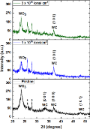
- , Netherlands) in the 2θ range from 20° to 80°. Scanning electron microscopy measurements of pristine and irradiated S1 films were performed using a MIRA Tescan II SEM at a high voltage of 25 kV using detector working in secondary-emission mode. Results and Discussion Phase and compositional studies of the
- samples underwent SEM measurements to see morphological changes under ion irradiation. The SEM images of the pristine S1 films and films irradiated at ion fluences of 1 × 10¹⁶ and 3 × 10¹⁶ ions/cm2 are shown in Figure 11. The SEM images clearly demonstrate significant morphological surface changes in the
- displacements results in pronounced surface restructuring. Similar phenomena, including ripple formation and surface roughening, have been widely reported and are known to depend on parameters such as ion energy, fluence, and target material properties [89][90]. The SEM and GIXRD results show strong correlation



Supramolecular one-dimensional conducting nanofibers from a C3-symmetric tetrathiafulvalene derivative
Beilstein J. Nanotechnol. 2026, 17, 872–881, doi:10.3762/bjnano.17.63
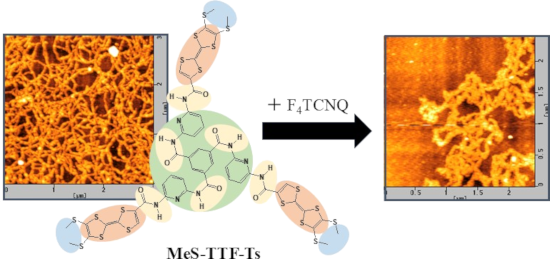
- structures with morphologies strongly dependent on the solvent conditions. AFM and SEM observations revealed that distinct supramolecular structures were formed depending on the solvent and substrate used, suggesting that molecular interactions and assembly processes play important roles in determining the
- marked differences in the observed morphologies. In addition to AFM observations, scanning electron microscopy (SEM) measurements were performed for representative samples deposited on HOPG substrates. Representative SEM images are provided in Supporting Information File 1 (Figures S2–S4). The
- morphologies observed by SEM differed from those obtained by AFM on mica substrates, showing more aggregated structures. This difference may originate from the distinct surface properties of the substrates used for the measurements. Although the detailed mechanism remains unclear, these results suggest that


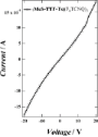
He+ FIBID-fabricated 3D AFM tip architectures: an exploratory study of hollow pillars, helices, and spirals
Beilstein J. Nanotechnol. 2026, 17, 863–871, doi:10.3762/bjnano.17.62
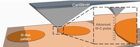
- observed variability among nominally similar structures and the post-operation changes documented by SEM, any apparent relation between geometry and AFM behaviour should be interpreted as qualitative and exploratory rather than conclusive. With these geometries established, we adopted a two-stage workflow
- step height values under matched conditions. In the second stage, the same tips were transferred to SEM in order to document their post-use condition. This before/after workflow allowed us to relate apparent AFM functionality to the actual post-operation morphology of the structures. From this point on
- , we focus on H2 and P5, which preserved their structural integrity through two AFM/SEM cycles, and on S7, which remained operational during the AFM measurements but was unavailable for the second SEM inspection because the cantilever was lost during subsequent SEM sample preparation (Figure 2). The


Glycerol photoelectrochemical oxidation reaction at carbon nitrides/BiVO4 materials
Beilstein J. Nanotechnol. 2026, 17, 806–817, doi:10.3762/bjnano.17.57
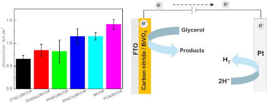
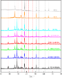
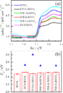
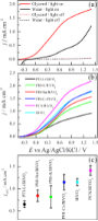
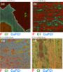
- spectroscopy (SEM-EDS) revealed regions rich in Bi and V, and others rich in C and N, suggesting the formation of heterojunctions. Photoelectrochemical studies demonstrated that BiVO4 is active for both water and glycerol (1.0 mol·L−1) oxidation, with a fourfold increase in photocurrent at 1.23 V vs reversible
- )/BiVO4. The SEM-EDS analysis after electrochemical tests revealed that the presence of crystalline CNs induces the segregation of vanadium oxides, contributing to a decrease in activity. On the other hand, the superior performance of PCN/BiVO4 is attributed to a greater thermal stability of PCN during
- acquired from 10° to 80° at a rate of 2°·min−1 with a step size of 0.02°. Morphological features and elemental composition were determined by field-emission scanning electron microscopy (FEG-SEM; Zeiss Supra35) coupled with energy-dispersive X-ray spectroscopy (EDS), operating at 15 kV. Optical properties




Restorative potential of laser-synthesized silver nanoparticles with Salvia officinalis for periodontal disease treatment: an in vitro study
Beilstein J. Nanotechnol. 2026, 17, 781–795, doi:10.3762/bjnano.17.55
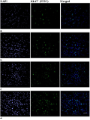
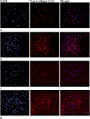
- presented as pg·mL−1 of IL-1β and IL-6. Statistical analysis For all performed assays, two independent experiments were performed, each in three replicas. The results are presented as mean value ± standard error of mean (SEM). Statistical analysis was performed using One-way ANOVA statistical test from SPSS



Tailoring Ag–Pt nanoalloys through solid-state dewetting: structural and optical insights
Beilstein J. Nanotechnol. 2026, 17, 748–759, doi:10.3762/bjnano.17.52

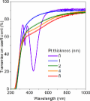
- a region with a lower film thickness at grain boundaries, which is well visible on the linear AFM profile in Figure 1 [45][46][47]. Figure 2a presents an exemplary scanning electron microscopy (SEM) image of nanostructures formed as a result of annealing of a silver–platinum bilayer with a thickness
- greater control over the final alloy composition. However, SEM observations revealed that an annealing temperature of 650 °C was insufficient to produce spherical, isolated nanostructures, instead resulting in the formation of irregularly shaped nanoislands. Therefore, the annealing temperature was
- gradually increased. At 700 °C, well-developed, isolated metallic islands with nearly spherical shapes were obtained, indicating a more advanced stage of morphological transformation. SEM images of the nanostructures formed after annealing at 650 °C, 675 °C, and 700 °C are presented in Figure 3a–c. In




Oxidative atmosphere-driven formation of single-phase spinel CuRh2O4 nanofibers for alkaline water oxidation
Beilstein J. Nanotechnol. 2026, 17, 737–743, doi:10.3762/bjnano.17.50
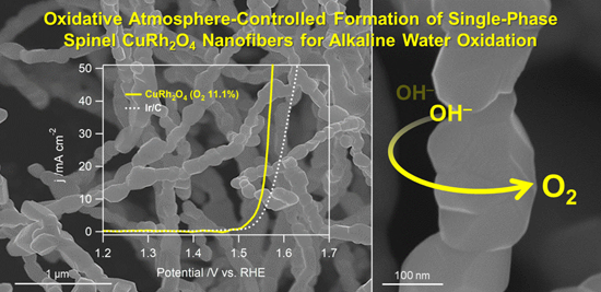
- electron microscopy (SEM) was employed to investigate the surface morphology and structural uniformity of the electrospun nanofibers after post-annealing. In the as-spun state prior to annealing, the fibers containing PVP exhibit smooth surfaces with an average diameter of approximately 300 nm (Supporting
- characterization Morphology and elemental composition of the synthesized nanomaterials were examined using field-emission scanning electron microscopy (FE-SEM; JEOL JSM-6700F) and high-resolution transmission electron microscopy (HRTEM; JEOL JEM-2100F). Surface chemical states and crystallographic structures were
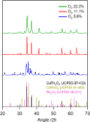
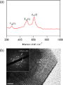
- nanomaterials annealed under the O2 concentration of 11.1% (inset: corresponding SAED pattern). SEM images of the as-prepared nanomaterials synthesized under O2 concentrations of (a) 5.6%, (b) 11.1%, and (c) 22.2%. (a) LSV polarization curves of the as-prepared Cu–Rh bimetallic oxides with commercial samples




Environmental applications of silver nanoparticles: state-of-the-art review and emerging trends
Beilstein J. Nanotechnol. 2026, 17, 697–736, doi:10.3762/bjnano.17.49
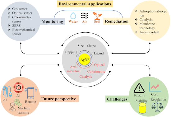
- X-ray analysis [49]. TEM analysis is performed on copper grids with varying mesh sizes and coatings, with carbon/formvar-coated copper grids commonly preferred. Scanning electron microscopy (SEM) provides information on the silver nanoparticles’ surface properties and the aggregation state of dried


Decontamination from water pollutants and pathogens by electrospun nanofibers doped with heavy-atom-free borafluorene-BODIPY photosensitizers
Beilstein J. Nanotechnol. 2026, 17, 668–682, doi:10.3762/bjnano.17.46
- microscopy (SEM) using a JSM-6010PLUS/LV InTouchScope™ system from JEOL (Tokyo, Japan), operating at an accelerating voltage of 11 kV. Prior to imaging, each nonwoven sample underwent a double-coating process with a thin gold layer (2–3 nm). Microstructural analysis was subsequently performed utilizing
- ) supported by Tukey’s post hoc test. Results and Discussion Selection of the matrix material Samples of BODIPY 1 encapsulated in poly(methyl methacrylate) (PMMA), polystyrene (PS), and polycaprolactone (PCL) nanofibers were formed using electrospinning, following by SEM analysis of the morphology. To ensure
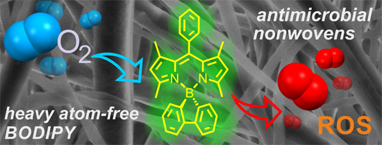
- good homogeneity, the initial amount of the photocatalyst 1 was set to 0.15 wt %. The SEM microstructures revealed fibers with uniform and smooth morphology (Figure 3). The comparison among 1(0.15 wt %)@PMMA, 1(0.15 wt %)@PS, and 1(0.15 wt %)@PCL revealed some differences in the average fiber diameter



Two-step laser synthesis of Ag@TiO2 nanomaterials for the photocatalytic degradation of rhodamine B
Beilstein J. Nanotechnol. 2026, 17, 622–634, doi:10.3762/bjnano.17.43
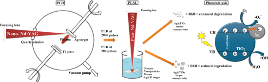
- characterization was performed using scanning electron microscopy (SEM) and transmission electron microscopy (TEM) with energy-dispersive spectroscopy (EDS), aiming to establish a clear correlation between synthesis parameters, nanostructure features, and photocatalytic activity. In our previous studies [6][7][21
- nanoparticle formation, leading to improved control over nanoparticle structure and photocatalytic performance. Results and Discussion SEM analysis The surface morphology of PLD-prepared samples was analyzed using SEM-EDS (Figure 2 and Figure 3). SEM images of the sample surface after deposition of silver on
- the titanium plate are given in Figure 2a and Figure 3a alongside the distribution of titanium (pink) and oxygen (blue), and silver (yellow) according to the elemental mapping obtained from the EDS analysis. SEM-SE images generated using a focused electron beam to image sample surfaces at high


Recent progress in enhancing built-in electric fields of perovskite solar cells via junction engineering
Beilstein J. Nanotechnol. 2026, 17, 602–621, doi:10.3762/bjnano.17.42
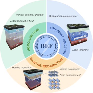
- , significantly lowering migration barriers and enhancing charge-separation efficiency. SEM and GIWAXS characterization of the SnAc2 + DMS-treated perovskite film is shown in Figure 2. Accordingly, the devices demonstrated exceptional performance, with carrier lifetimes extended to 812 ns and a PCE increased to



Synthesis of Cu–Mo/TiO2 and Co–Mo/TiO2 photocatalysts for the efficient degradation of organic pollutants in water
Beilstein J. Nanotechnol. 2026, 17, 559–570, doi:10.3762/bjnano.17.37
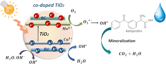
- , copper and cobalt, were added at 0.2–0.5 wt %. The co-doped TiO2 photocatalysts were characterized by XRD, SEM, N2 physisorption, UV–vis diffuse reflectance spectroscopy, and photoluminescence spectroscopy. The structural characterization showed stabilization of the anatase phase, and lattice distortion
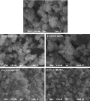
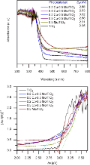
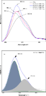
- patterns of TiO2 and co-doped TiO2 materials and (b) diffraction peak shift in TiO2 materials. SEM images of TiO2 and co-doped TiO2 materials. Diffuse reflectance UV–vis spectra of TiO2 and co-doped TiO2 materials. (a) Photoluminescence spectra of TiO2 and co-doped TiO2 materials and (b) deconvoluted



Electrochemical determination of ciprofloxacin using a MIL-101/reduced graphene oxide-modified electrode
Beilstein J. Nanotechnol. 2026, 17, 541–554, doi:10.3762/bjnano.17.35
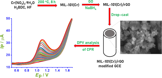
- integration of MIL-101(Cr) and rGO, while preserving the Cr3+ oxidation state and the characteristic coordination environment of the MIL-101 framework. The SEM images of rGO, MIL-101, and the MIL-101/rGO composite are shown in Figure 4. The rGO sample (Figure 4a) displays a typical wrinkled, sheet-like
- mapping, and (e) carbon element mapping. XPS spectra of the MIL-101/rGO (3.3): (a) survey spectrum, (b) high-resolution Cr 2p spectrum, (c) high-resolution C 1s spectrum, and (d) high-resolution O 1s spectrum. SEM observation of (a) rGO, (b) MIL-101, and (c) MIL-101(Cr)/rGO (3.3). (a) Cyclic voltammograms


Defects and defect-mediated engineering of two-dimensional materials: challenges and open questions
Beilstein J. Nanotechnol. 2026, 17, 454–488, doi:10.3762/bjnano.17.31





Eco-efficient materials for agricultural crops based on a mineral rich in MOR- and HEU-type zeolites
Beilstein J. Nanotechnol. 2026, 17, 381–395, doi:10.3762/bjnano.17.26
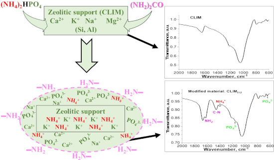
- microscopy (SEM), scanning transmission electron microscopy (STEM), N2 physisorption, and other research methods. Results and Discussion Characterization of DAP-modified zeolite CLIM The chemical compositions of natural zeolite (CLIM) from the San Andrés deposit and its modified forms (CLIMf) with ammonium
- these experimental materials, studies were conducted using XRD, N2 adsorption isotherms, SEM, TEM, and STEM. However, they will only be presented for the most interesting materials, namely those that were modified with 2.0% and 3.5% DAP and then with a 5 M urea solution, that is, the materials CLIMf2
- , some with a fibrous shape (as shown in Figure 5) and others acicular (as observed more clearly in Figure 6b) [45][46]. HEU crystals with laminar habit can also be distinguished, the morphology of which is associated with the presence of potassium [43][44]. The SEM images (Figure 6) of the modified




Polycatecholamine nanocoatings on stainless steel: the effect on attachment of human fibroblasts and platelets
Beilstein J. Nanotechnol. 2026, 17, 365–380, doi:10.3762/bjnano.17.25
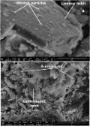
- times were subjected for further tests, that is, PDA 10 min, PDA 4 h, PTYR 30 min, and PTYR 24 h. Thickness of the coatings Thickness of the coatings was measured with SEM thanks to the tilt of the samples in the scratch location (Figure 3). The average thicknesses of PDA 10 min and 4 h coatings were
- longest time of coating. Images taken directly after coating (Figure 5a,b,e,f) show the initial coating morphology. The surface of SS-PDA 10 min could not be clearly distinguished with SEM; however, the surface of SS-PDA 4 h presented a full coverage with a polymer film. After 28 d of constant washing
- with PBS, the coating is still present on the SS-PDA 4 h variant, although it seems to be cracked. The coating presence on the SS-PDA 10 min variant could not be confirmed with SEM after 28 d of washing. The morphology of PTYR nanocoatings was significantly different. In Figure 5e,f, bright micro




Interconnection morphology effects on the radio frequency response of carbon nanotube sponges
Beilstein J. Nanotechnol. 2026, 17, 343–351, doi:10.3762/bjnano.17.23
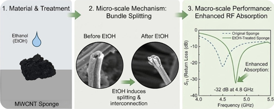
- findings point to a different interpretation related to the morphological effect of ethanol on the CNT mesh [27][29]. A scanning electron microscopy (SEM) study provided insights into the microstructure of the CNT film (Supporting Information File 1, Figure S1) and the sponges (Figure 3). The study
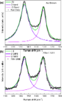
- interconnected structure of the samples. The Raman analysis results are in line with the morphological information obtained from the SEM data. The SEM and Raman experiments confirm the effect of ethanol on the CNS, which improves the presence of grooves and pores as available adsorption sites after ethanol
- fits in Figure 5. From the Raman and XPS studies, as well as the SEM micrographs, we conclude that the ethanol treatment induced a change in the structure of the CNS sample, unravelling many of the aggregated tubes, resulting in an increase in pore size and overall available surface area. Without



Beam shaping techniques for pulsed laser ablation in liquids: Unlocking tunable control of nanoparticle synthesis in liquids
Beilstein J. Nanotechnol. 2026, 17, 309–342, doi:10.3762/bjnano.17.22
- rates, thereby limiting coalescence and favouring the formation of smaller NPs. Figure 13 compares SEM images and size distributions of Au NPs produced by PLAL in water with both laser beams. SEM analysis of Au NPs revealed that DS pulses suppressed aggregation, eliminating large particles, unlike







Advancing nanolithography: a comprehensive review of materials for local anodic oxidation with AFM
Beilstein J. Nanotechnol. 2026, 17, 275–291, doi:10.3762/bjnano.17.19


Durable antimicrobial activity of fabrics functionalized with zeolite ion-exchanged nanomaterials against Staphylococcus aureus and Escherichia coli
Beilstein J. Nanotechnol. 2026, 17, 262–274, doi:10.3762/bjnano.17.18
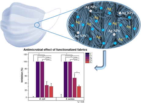
- of copper and zinc in the functionalized textiles analyzed by EDS was nearly four times lower in comparison with Ag/CBV-600-bramante. This may be explained by non-homogeneous loading of nanomaterials on the textile, as also evidenced by SEM images. The Si/Al molar ratio for Y zeolite was 2.7, which
- impurities in the samples were found. SEM analysis Figure 2 shows the micrographs of bramante fabric before and after its functionalization with 0.5 g of Ag/CBV-600, Cu/CBV-600, and Zn/CBV-600 nanomaterials using the pad–dry–cure method. As seen in Figure 2a, the original fabric did not show fiber
- after four wash cycles. Such findings are significant for the potential applications of these fabrics in the manufacture of personal protective equipment. SEM-EDS analysis of washed fabrics To further evaluate the durability of the functionalized fabrics under practical conditions, the samples underwent




Multilayered hyperbolic Au/TiO2 nanostructures for enhancing the nonlinear response around the epsilon-near-zero point
Beilstein J. Nanotechnol. 2026, 17, 251–261, doi:10.3762/bjnano.17.17
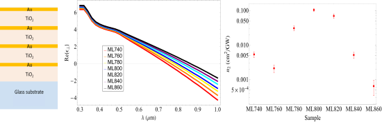
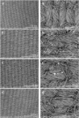
- the deposited layers. In Table 1, we can see that we have widths for every stack that are different from the ones proposed for the simulated ENZ points. This change in thickness was also seen in scanning electron microscopy (SEM). In Figure 4a we present a SEM image of the ML800 structure; we see that


Comparative study on 3D morphologies of delignified, single tracheids and fibers of five wood species
Beilstein J. Nanotechnol. 2026, 17, 239–250, doi:10.3762/bjnano.17.16
- nitrogen and subsequent lyophilization overnight (−45 °C, 0.080 mbar, Christ Alpha 2–4 LDplus, Christ, Osterode, Germany). Scanning electron microscopy Air-dried wood samples of each of the five species (untreated and delignified) were glued onto metal (SEM) stubs and sputter-coated with a 10 nm layer of
- gold–palladium (Leica Bal-TEC SCD500, Leica, Wetzlar, Germany). The samples were then visualized using a scanning electron microscope (SEM) Hitachi S-4800 (Hitachi, Tokyo, Japan) at an accelerating voltage of 1.5 kV. Nanotomography and 3D reconstruction Single fibers and tracheids were easily
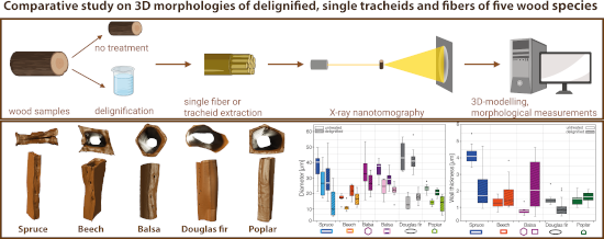
- size resulted from the quality of the scans, as some scans were not of sufficient quality for the purpose of this study. We therefore only worked with the scans with sufficient quality (16 in total). Moreover, the diameter and wall thickness were additionally measured from SEM images of the untreated


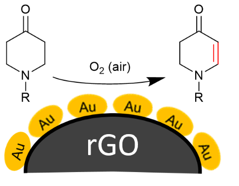
Gold nanoparticle-decorated reduced graphene oxide as a highly effective catalyst for the selective α,β-dehydrogenation of N-alkyl-4-piperidones
Beilstein J. Nanotechnol. 2026, 17, 218–238, doi:10.3762/bjnano.17.15
- , scanning electron microscopy (SEM) and nitrogen gas sorption surface area analysis using the Brunauer–Emmett–Teller (BET) theory were employed to evaluate the surface structure, porosity, and overall texture of the materials. The SEM image in Figure 2a reveals the typical structure of AC, characterized by
- small particles. The nitrogen sorption isotherms and their hysteresis loops in Figure 3 supplement the SEM analysis of the three carbon materials in terms of specific BET surface area, and pore volume and size. AC features a reversible almost type-I isotherm, given by microporous materials with narrow


Time of flight secondary ion mass spectrometry imaging of contaminant species in chemical vapour deposited graphene on copper
Beilstein J. Nanotechnol. 2026, 17, 200–213, doi:10.3762/bjnano.17.13
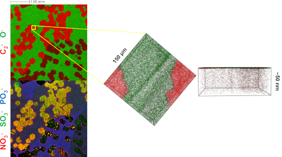
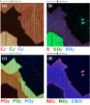
- a Wyko NT1100 White Light Optical Profiling System using a 20× magnification in vertical scanning interferometry (VSI) mode. Scanning electron microscopy (SEM) was carried out with a Carl Zeiss SIGMA VP at an acceleration voltage of 2 kV to ensure sample consistency. Four separate Cu foil samples





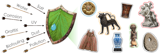
Functional surface engineering for cultural heritage protection: the role of superhydrophobic and superoleophobic coatings – a comprehensive review
Beilstein J. Nanotechnol. 2026, 17, 63–96, doi:10.3762/bjnano.17.6