
Search results
Search for "transmission electron microscopy" in Full Text gives 704 result(s) in Beilstein Journal of Nanotechnology. Showing first 200.
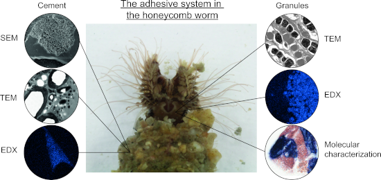
The cement of the tube-dwelling polychaete Sabellaria alveolata: a complex composite adhesive material
Beilstein J. Nanotechnol. 2025, 16, 1998–2014, doi:10.3762/bjnano.16.138
- ) equipped with an 80 mm2 silicon drift detector. Acquisition conditions on the SEM were 15 kV, 10 mm working distance, and 10 s live time acquisition at approximately 30–40% dead time. The spectra were acquired with an AZtec (Oxford Instrument) EDS data processing software. Transmission electron microscopy
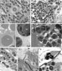
- building organ form the external part of a complex secretory organ made up of clusters of cement cells located deep within the parathoracic segments of the worm [6][17]. Using transmission electron microscopy, two main types of cement cells can be distinguished based on the ultrastructure of their



Laser ablation in liquids for shape-tailored synthesis of nanomaterials: status and challenges
Beilstein J. Nanotechnol. 2025, 16, 1963–1997, doi:10.3762/bjnano.16.137
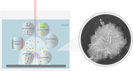
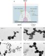
- observed the formation of a mixture of nanosheets and spherical NPs for the ablation using the 1064 nm laser wavelength, while at 532 nm only spherical NPs were observed. The authors observed nanosheets only in scanning electron microscopy, while transmission electron microscopy showed only spherical




Self-assembly and adhesive properties of Pollicipes pollicipes barnacle cement protein cp19k: influence of pH and ionic strength
Beilstein J. Nanotechnol. 2025, 16, 1863–1872, doi:10.3762/bjnano.16.129
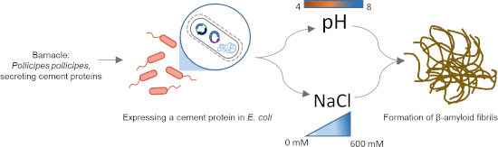
- bioinspired adhesives and biomaterials. Keywords: adhesive; amyloid fibre; barnacle cement protein; surface coating; transmission electron microscopy; Introduction Marine adhesives are naturally occurring substances secreted by a variety of organisms to attach themselves to submerged surfaces such as rocks
- work, we expressed recombinant P. pollicipes cp19k (rPpolcp19k) in E. coli and identified key environmental modulators of fibril formation by the protein. Transmission electron microscopy (TEM) was used to study the rate of fibril formation and morphology under varied pH and salt concentration
- achieved (Supporting Information File 1, Figure S1). Purified protein was dialysed into 10 mM sodium acetate (pH 4.0) or 10 mM sodium phosphate (pH 8.0) buffer, with NaCl concentrations ranging from 0 to 600 mM. Transmission electron microscopy TEM analysis identified the formation of fibril structures by


Phytol-loaded soybean oil nanoemulsion as a promising alternative against Leishmania amazonensis
Beilstein J. Nanotechnol. 2025, 16, 1826–1836, doi:10.3762/bjnano.16.126
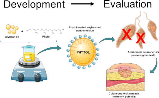
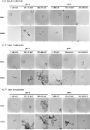
- shown in the size distribution graph (Figure 1b). Moreover, the zeta potential (ZP) of both samples was around −20 mV, with no significant changes upon phytol loading. To evaluate the shape and morphological characteristics of the nanoemulsions, transmission electron microscopy (TEM) analysis was
- evaluated over a 30-day period by monitoring droplet size, PdI, ZP, and pH using the same methodologies previously described. Morphology The morphology of the nanoemulsion droplets was examined by transmission electron microscopy using a FEI Tecnai G2 Spirit Biotwin microscope operating at 120 kV (FEI


Exploring the potential of polymers: advancements in oral nanocarrier technology
Beilstein J. Nanotechnol. 2025, 16, 1751–1793, doi:10.3762/bjnano.16.122



Multifunctional anionic nanoemulsion with linseed oil and lecithin: a preliminary approach for dry eye disease
Beilstein J. Nanotechnol. 2025, 16, 1711–1733, doi:10.3762/bjnano.16.120

- digital pH meter (HI2221, Hanna Instruments, BR) equipped with a calibrated glass electrode and a temperature sensor. The electrode and sensor were immersed in the samples, and pH readings were recorded once the measurements stabilized. Transmission electron microscopy The morphology of the nanoemulsions
- was analyzed using transmission electron microscopy (TEM) with a MORGAGNI 268D (FEI Company, USA), operated at 80 kV. Before imaging, the samples were sonicated in an ultrasound bath for 15 min, and a drop of the suspension was placed onto a copper grid (200 mesh) coated with formvar/carbon. Excess



Prospects of nanotechnology and natural products for cancer and immunotherapy
Beilstein J. Nanotechnol. 2025, 16, 1644–1667, doi:10.3762/bjnano.16.116

- the development of reactive oxygen species (ROS) [118]. Hyaluronic acid can inhibit local recurrence and distant tumor growth when used in immunotherapy scenarios in conjuction with CAR-T cells and anti-PDL1-conjugated platelets [119]. Transmission electron microscopy (TEM) and dynamic light



Photocatalytic degradation of ofloxacin in water assisted by TiO2 nanowires on carbon cloth: contributions of H2O2 addition and substrate absorbability
Beilstein J. Nanotechnol. 2025, 16, 1567–1579, doi:10.3762/bjnano.16.111
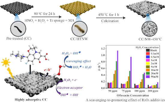
- drying. The P25 loading was ca. 50.0 mg (≈1.0 mg/cm2), nearly the same as that of the TiO2 nanowires on carbon cloth. Characterizations The morphologies were analyzed using field-emission scanning electron microscopy (FESEM, Zeiss Germany Sigma 300) and high-resolution transmission electron microscopy


Influence of laser beam profile on morphology and optical properties of silicon nanoparticles formed by laser ablation in liquid
Beilstein J. Nanotechnol. 2025, 16, 1533–1544, doi:10.3762/bjnano.16.108
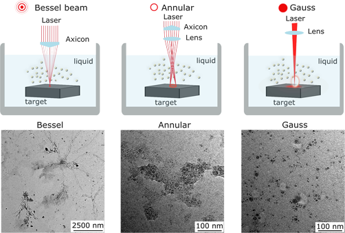
- ultraviolet–visible spectroscopy, high-resolution transmission electron microscopy, and Raman and photoluminescence spectroscopies, and the correlation of the NP properties with the laser beam profile was studied. Three different beam profiles were selected, namely, a Bessel beam produced using an axicon, an
- structure of the formed NPs were analyzed by means of ultraviolet–visible (UV–vis) spectroscopy, high-resolution transmission electron microscopy (HRTEM), selected area electron diffraction (SAED), X-ray diffraction (XRD), and Raman and photoluminescence (PL) spectroscopies. Particle size and morphology
- were investigated by transmission electron microscopy (TEM) using a JEOL JEM-2100F (JEOL, USA) operating with an accelerating voltage of 200 kV. For TEM and SAED measurements, the NPs were drop-casted onto copper grids covered by carbon film and dried under ambient conditions. Crystalline structure


Laser processing in liquids: insights into nanocolloid generation and thin film integration for energy, photonic, and sensing applications
Beilstein J. Nanotechnol. 2025, 16, 1428–1498, doi:10.3762/bjnano.16.104
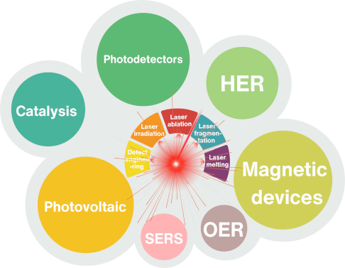
- films of copper phthalocyanine (CuPc) doped with laser generated Au and Ag NPs were prepared using spin coating on glass substrates. Transmission electron microscopy analysis indicated spherical NPs having sizes in the range of 7–43 nm for Au NPs, and 12–50 nm for Ag NPs. The deposited films had good






Photochemical synthesis of silver nanoprisms via green LED irradiation and evaluation of SERS activity
Beilstein J. Nanotechnol. 2025, 16, 1417–1427, doi:10.3762/bjnano.16.103
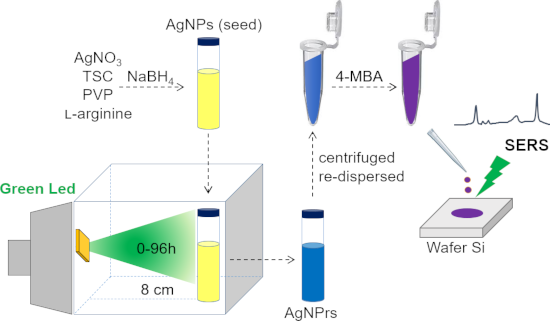
- ± 20 nm) for various periods of time up to 72 h. The growth mechanism was investigated through ultraviolet–visible spectroscopy, field-emission scanning electron microscopy, X-ray diffraction, and transmission electron microscopy analyses, confirming the gradual transformation of spherical seeds into
- nm. Samples were diluted tenfold with deionized water prior to measurement. To determine the morphology, particle size of the synthesized nanostructures, the samples were analyzed using field-emission scanning electron microscopy (FESEM, Hitachi S-4800, Japan) and transmission electron microscopy


Ferroptosis induction by engineered liposomes for enhanced tumor therapy
Beilstein J. Nanotechnol. 2025, 16, 1325–1349, doi:10.3762/bjnano.16.97
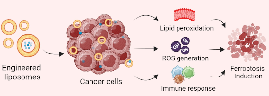
- light scattering (DLS) is commonly used to determine liposome size and size distribution. Transmission electron microscopy (TEM) and atomic force microscopy (AFM) can be used to image liposome morphology and determine lamellarity. Zeta potential measurements assess the surface charge of liposomes, which


Deep-learning recognition and tracking of individual nanotubes in low-contrast microscopy videos
Beilstein J. Nanotechnol. 2025, 16, 1316–1324, doi:10.3762/bjnano.16.96
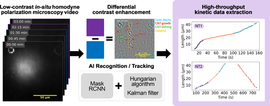
- situ imaging platforms for nanomaterials, such as environmental transmission electron microscopy (ETEM) of CNTs [31][33] and environmental scanning electron microscopy (ESEM) of graphene growth and etching processes [34]. Snapshots from (a) the raw video of carbon nanotube (CNT) synthesis on a stable
- , finding numerous applications in scientific research. Deep learning models have been used to identify two-dimensional materials in microscopic images [6], characterize mineral composition in scanning electron microscopy (SEM) samples [7], and determine nanotube chirality from transmission electron
- microscopy images [8]. Here, we report on using a deep learning algorithm to automatically recognize and track individual carbon nanotubes during growth, captured in polarization microscopy video sequences. Materials and Methods CNT growth The experimental setup was previously described in [4][9]. In short


Enhancing the photoelectrochemical performance of BiOI-derived BiVO4 films by controlled-intensity current electrodeposition
Beilstein J. Nanotechnol. 2025, 16, 1289–1301, doi:10.3762/bjnano.16.94
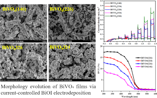
- ) was employed to determine the chemical states of each photoanode. All photoanode morphologies were examined using field-emission transmission electron microscopy (FESEM, Hitachi SU8010). Photoelectrochemical measurements PEC experiments were performed in a conventional three-electrode cell using an



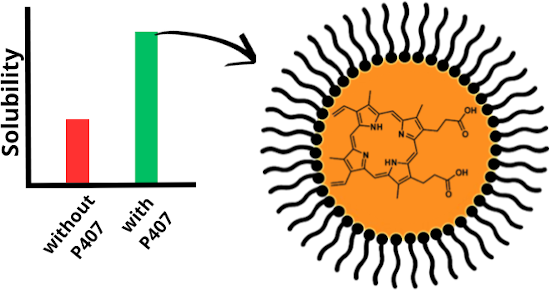
Investigation of the solubility of protoporphyrin IX in aqueous and hydroalcoholic solvent systems
Beilstein J. Nanotechnol. 2025, 16, 1209–1215, doi:10.3762/bjnano.16.89
- using a previously validated method, yielding a correlation coefficient R = 0.9973 [20][21]. Transmission electron microscopy Transmission electron microscopy (TEM) analysis was carried out using a JEM-1400 microscope (JEOL, Tokyo, Japan) with an accelerating voltage of 100 kV. The samples were diluted


Chitosan nanocomposite containing rotenoids: an alternative bioinsecticidal approach for the management of Aedes aegypti
Beilstein J. Nanotechnol. 2025, 16, 1197–1208, doi:10.3762/bjnano.16.88
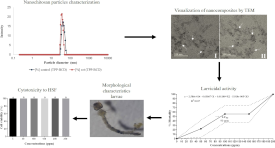
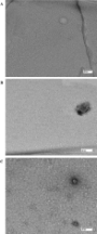
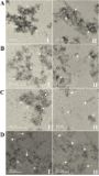
- (control = empty particles; rot = rotenoid-loaded particles). B) Controlled ionic gelation of CS with TPP-β-CD inclusion complex (control and rot). Transmission electron microscopy (TEM) of chitosan nanocomposites. A) Conventional ionic gelation of CS/TPP. B) Conventional ionic gelation of CS/TPP



Crystalline and amorphous structure selectivity of ignoble high-entropy alloy nanoparticles during laser ablation in organic liquids is set by pulse duration
Beilstein J. Nanotechnol. 2025, 16, 1141–1159, doi:10.3762/bjnano.16.84
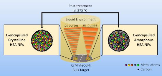
- in organic solvents (acetone, ethanol, acetonitrile). In a systematic experimental series using high-resolution transmission electron microscopy, scanning transmission electron microscopy with energy-dispersive X-ray spectroscopy, selected-area electron diffraction, X-ray diffraction, electron energy
- heterogeneous catalysts or magnets. In that context, the outstanding temperature stability up to 375 °C (differential scanning calorimetry) or 500 °C (transmission electron microscopy) may motivate future application-relevant work. Keywords: amorphous; cantor alloy; compositionally complex alloy; complex solid
- . Nanoparticle characterization is done by high-resolution transmission electron microscopy (HRTEM), energy-dispersive X-ray spectroscopy (STEM-EDX), selected-area electron diffraction (SAED), X-ray diffraction (XRD), and electron energy loss spectroscopy (EELS), complemented by tempering and laser post




Towards a quantitative theory for transmission X-ray microscopy
Beilstein J. Nanotechnol. 2025, 16, 1113–1128, doi:10.3762/bjnano.16.82
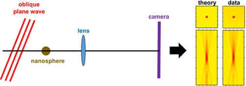
- (Supporting Information File 1, Section S12) and the non-tilted condenser model was much easier to compute. To enable a more precise quantitative analysis, we first extensively characterized the gold nanospheres by both scanning and transmission electron microscopy (SEM and TEM, Supporting Information File 1



Piezoelectricity of hexagonal boron nitrides improves bone tissue generation as tested on osteoblasts
Beilstein J. Nanotechnol. 2025, 16, 1068–1081, doi:10.3762/bjnano.16.78
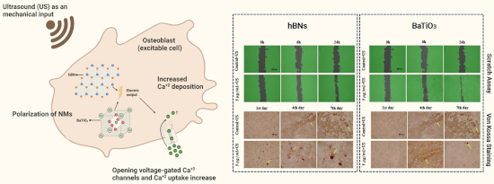
- SiC surface and stored at room temperature. The synthesized hBN and commercial BaTiO3 nanoparticles (Sigma-Aldrich, Germany) were characterized using several analytical techniques. Transmission electron microscopy (TEM, JEOL ARM 200 CF, 200 keV) was employed to examine morphology and particle size by


Structural and magnetic properties of microwave-synthesized reduced graphene oxide/VO2/Fe2O3 nanocomposite
Beilstein J. Nanotechnol. 2025, 16, 921–932, doi:10.3762/bjnano.16.70
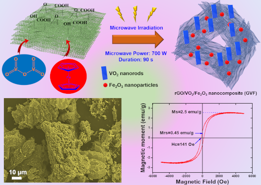
- and 6.50 mA using an Al Kα. The morphologies and elemental analyses of rGO and the NCs were analyzed through scanning electron microscopy (SEM, Hitachi, S-4800). The structural analysis of these fabricated NCs was examined using high-resolution transmission electron microscopy (HRTEM, FEI Tecnai G2



Focused ion beam-induced platinum deposition with a low-temperature cesium ion source
Beilstein J. Nanotechnol. 2025, 16, 910–920, doi:10.3762/bjnano.16.69
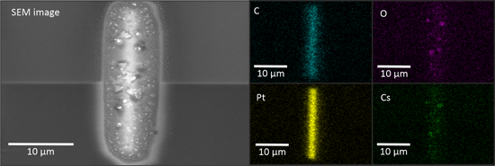
- energy-dispersive X-ray spectroscopy (EDS), lamellas for transmission electron microscopy (TEM) were prepared. Experimental The Ga+ FIB is a ThermoFisher Helios NanoLab 650 and uses a gas injection system (GIS). ZeroK NanoTech Corporation has created commercially available Cs+ FIB systems based on


Heat-induced transformation of nickel-coated polycrystalline diamond film studied in situ by XPS and NEXAFS
Beilstein J. Nanotechnol. 2025, 16, 887–898, doi:10.3762/bjnano.16.67
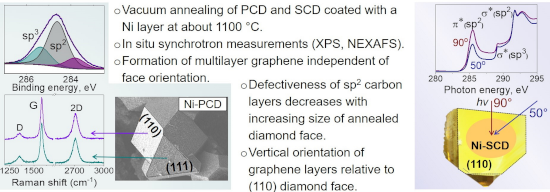
- process occurring at the interface between diamond and Ni nanoparticles was revealed using high-resolution transmission electron microscopy (HRTEM) [19][24]. During annealing, Ni nanoparticles etch the diamond surface, resulting in the formation of a narrow interdiffusion zone. The carbon atoms released


Synthesis and magnetic transitions of rare-earth-free Fe–Mn–Ni–Si-based compositionally complex alloys at bulk and nanoscale
Beilstein J. Nanotechnol. 2025, 16, 823–836, doi:10.3762/bjnano.16.62
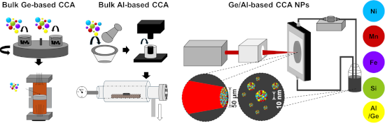
- 2.8 J·cm−2 [41]. Additionally, to increase the NP concentration and reduce the organic solvent use, a closed loop liquid flow system was employed. Size distribution, morphology, elemental composition, and crystal structure of the NPs from both CCA targets were examined using transmission electron
- microscopy (TEM) and EDX with a Jeol 2200FS microscope (Japan) equipped with an Oxford X-MaxN TLE 80 EDX detector (UK). The microscope was operated at an acceleration voltage of 200 kV and utilized a 2k × 2k GATAN UltraScan 1000XP CCD camera. For TEM analysis, the colloidal particles were dispersed onto a


Morphology and properties of pyrite nanoparticles obtained by pulsed laser ablation in liquid and thin films for photodetection
Beilstein J. Nanotechnol. 2025, 16, 785–805, doi:10.3762/bjnano.16.60
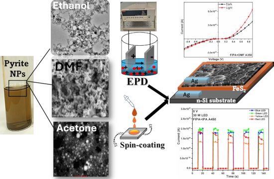
- nanocolloid in this work. The optical properties of nanocolloids and their thin films were evaluated using UV–visible (UV–vis) spectroscopy. The nanoparticle characterization and surface morphology were studied using transmission electron microscopy (TEM), scanning electron microscopy (SEM), and the
- temperatures and amounts of sulfur. Characterization The morphological analyses of pyrite NPs were recorded using the FEI Titan G2 80–300 for TEM, high-angle annular dark-field scanning transmission electron microscopy (HAADF-STEM), high-resolution TEM (HRTEM) and selected area electron diffraction (SAED). The
- disulfide (FeS2)) nanoparticles (NPs) of different morphologies using pulsed laser ablation in liquid (PLAL) in different organic solvents. The impact of the solvent on the morphological, compositional, and optical properties of the synthesized NPs is investigated by techniques such as transmission electron




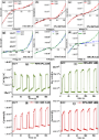


Changes of structural, magnetic and spectroscopic properties of microencapsulated iron sucrose nanoparticles in saline
Beilstein J. Nanotechnol. 2025, 16, 762–784, doi:10.3762/bjnano.16.59
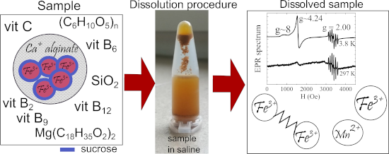
- changes upon dissolution in saline were tested. For the undissolved sample, calcium alginate microcapsules with irregular shapes were registered via scanning electron microscopy, inside which core–shell nanoparticles were identified by transmission electron microscopy micrographs. Magnetic studies (DC and
- transmission electron microscopy (TEM). Composition studies using XRD, magnetic properties using dc and ac magnetometry, and extensive spectral analysis using Fourier-transform infrared spectroscopy (FTIR), Raman, and electron paramagnetic resonance (EPR) were also performed. Considering that the AB-Fortis
- nm, cores, while the thickness of the shell seems constant ≈10 nm. The sucrose shell is homogenous; thus the individual cores appear to be well separated from each other. In Figure 3c, the high-resolution transmission electron microscopy (HRTEM) image presents a single crystalline nanoparticle. The